The RSNA Abdominal Traumatic Injury CT (RATIC) Dataset Authors: Jeffrey D. Rudie, Hui-Ming Lin, Robyn L. Ball, Sabeena Jalal, Luciano M. Prevedello, Savvas Nicolaou, Brett S. Marinelli, Adam E. Flanders, Kirti Magudia, George Shih, Melissa A.
Davis, John Mongan, Peter D. Chang, Ferco H. Berger, Sebastiaan Hermans, Meng Law, Tyler Richards, Jan-Peter Grunz, Andreas Steven Kunz, Shobhit Mathur, Sandro Galea-Soler, Andrew D. Chung, Saif Afat, Chin-Chi Kuo, Layal Aweidah, Ana Villanueva Campos, Arjuna Somasundaram, Felipe Antonio Sanchez Tijmes, Attaporn Jantarangkoon, Leonardo Kayat Bittencourt, Michael Brassil, Ayoub El Hajjami, Hakan Dogan, Muris Becircic, Agrahara G.
Bharatkumar, Eduardo Moreno Júdice de Mattos Farina, Dataset Curator Group, Dataset Contributor Group, Dataset Annotator Group, Errol Colak.
2
Liverpool Hospital, Sydney, Australia (L.A.), Hospital Universitario Ramón y Cajal, Madrid, Spain Diagnostic Imaging, Universidade Federal de São Paulo, São Paulo, Brazil (E.M.J.M.F.),
Dataset Curator Group:
Matthew Aitken, Patrick Chun-Yin Lai, Priscila Crivellaro, Jayashree Kalpathy-Cramer, Zixuan Hu, Reem Mimish, Aeman Muneeb, Mitra Naseri, Maryam Vazirabad, Rachit Saluja
Dataset Contributor Group:
Nitamar Abdala, Jason Adleberg, Waqas Ahmad, Christopher O. Ajala, Emre Altinmakas, Robin Ausman, Miguel Ángel Gómez Bermejo, Deniz Bulja, Jeddi Chaimaa, Lin-Hung Chen, Sheng- Hsuan Chen, Hsiu-Yin Chiang, Rahin Chowdhury, David Dreizin, Zahi Fayad, Yigal Frank, Sirui Jiang, Belma Kadic, Helen Kavnoudias, Alexander Kagen, Felipe C. Kitamura, Nedim Kruscica, Michael Kushdilian, Brian Lee, Jennifer Lee, Robin Lee, Che-Chen Lin, Karun Motupally, Eamonn Navin, Andrew S. Nencka, Christopher Newman, Akdi Khaoula, Shady Osman, William Parker, Jacob J. Peoples, Marco Pereañez, Christopher Rushton, Navee Sidi Mahmoud, Xueyan Mei, Beverly Rosipko, Muhammad Danish Sarfarz, Adnan Sheikh, Maryam Shekarforoush, Amber Simpson, Ashlesha Udare, Victoria Uram, Emily V. Ward, Conor Waters, Min-Yen Wu, Wanat
Dataset Annotator Group:
Claire K. Sandstrom, Angel Ramon Sosa Fleitas, Joel Kosowan, Christopher J Welman, Sevtap Arslan, Mark Bernstein, Linda C. Chu, Karen S. Lee, Chinmay Kulkarni, Taejin Min, Ludo Beenen, Betsy Jacobs, Scott Steenburg, Sree Harsha Tirumani, Eric Wallace, Shabnam Fidvi, Helen Oliver, Casey Rhodes, Paulo Alberto Flejder, Adnan Sheikh, Muhammad Munshi, Jonathan Revels, Vinu Mathew, Marcela De La Hoz Polo, Apurva Bonde, Ali Babaei Jandaghi, Robert Moreland, M. Zak Rajput, James T. Lee, Nikhil Madhuripan, Ahmed Sobieh, Bruno Nagel Calado, Jeffrey D Jaskolka, Lee Myers, Laura Kohl, Matthew Wu, Wesley Chan, Facundo Nahuel Diaz
Abstract:
The RSNA Abdominal Traumatic Injury CT (RATIC) dataset is the largest publicly available collection of adult abdominal CT studies annotated for traumatic injuries. This dataset includes 4,274 studies from 23 institutions across 14 countries. The dataset is freely available for non- commercial use via Kaggle at https://www.kaggle.com/competitions/rsna-2023-abdominal- trauma-detection. Created for the RSNA 2023 Abdominal Trauma Detection competition, the dataset encourages the development of advanced machine learning models for detecting abdominal injuries on CT scans. The dataset encompasses detection and classification of traumatic injuries across multiple organs, including the liver, spleen, kidneys, bowel, and mesentery. Annotations were created by expert radiologists from the American Society of Emergency Radiology (ASER) and Society of Abdominal Radiology (SAR). The dataset is annotated at multiple levels, including the presence of injuries in three solid organs with injury grading, image-level annotations for active extravasations and bowel injury, and voxelwise segmentations of each of the potentially injured organs. With the release of this dataset, we hope to facilitate research and development in machine learning and abdominal trauma that can lead to improved patient care and outcomes.

Introduction
Trauma is the most common cause of fatal injuries in Americans under the age of 45 and claims six million lives globally each year (1). Early accurate diagnosis and grading of traumatic injuries is critical in guiding clinical management and improving patient outcomes. Computed tomography plays a central role in the initial evaluation of hemodynamically stable patients (2,3).
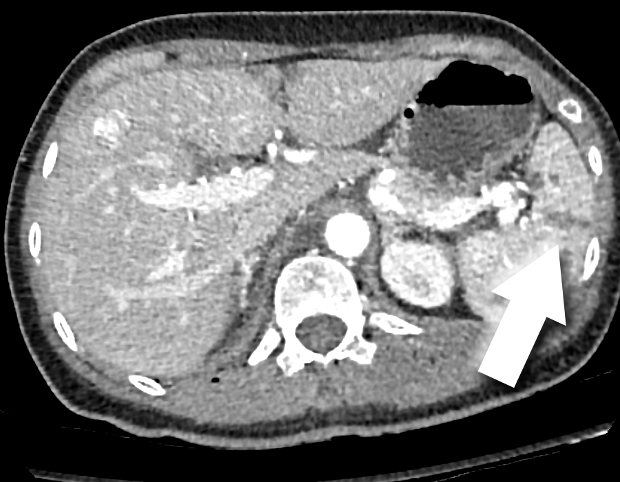
For blunt and penetrating abdominal trauma, the American Association for the Surgery of Trauma (AAST) organ injury grading system is the most well recognized system for grading solid organ injuries (4,5) and is critical in triaging patients between surgery, minimally invasive intervention, and conservative management (6).
While the AAST grading system is an important guide to assess solid organ injury, rapid interpretation of trauma studies is challenging given the large number of images needed to review and potential for subtle findings. In fact, diagnostic errors in the interpretation of trauma are common (7) and there is high inter-rater variability in the AAST grading system (8,9). Furthermore, the large variation in protocols used at different hospitals, including a single portal venous phase, multiphasic imaging, and split bolus approaches (10), can further complicate this task.
Automated assessment of traumatic abdominal injuries is an excellent use case for artificial intelligence (AI) algorithms given the potential to prioritize studies that may require more expedient interpretations as well as augment radiologist accuracy and efficiency, which may be particularly true in areas where subspecialists are in short supply. Recent work on AI based assessment of abdominal trauma includes studies on automated detection of splenic (11-14) and liver (15) injury, hemoperitoneum (16), and pneumoperitoneum (17). However, prior studies have typically been limited in scope to single organs and single institutions, and realistically not generalizable into clinical practice. Thus, there is a need for large multi-institutional publicly available annotated abdominal trauma datasets to address this challenge.
6
The Radiological Society of North America (RSNA) collaborated with the American Society of Emergency Radiology (ASER) and Society of Abdominal Radiology (SAR) to curate a large, publicly available expert-labeled dataset of abdominal CT images for traumatic injuries focusing on injuries to the liver, spleen, kidneys, bowel and mesentery, and active extravasation. This dataset was used for the RSNA 2023 Abdominal Trauma Detection competition which attracted 1,500 competitors from around the world to develop innovative machine learning (ML) models that detect traumatic injuries on abdominal CT.
Dataset Description And Usage
The RSNA Abdominal Traumatic Injury CT (RATIC) dataset is composed of CT scans of the abdomen and pelvis from 4,274 patients with a total of 6,481 image series from 23 institutions across 14 countries and 6 continents. A detailed breakdown of patient demographics and injuries across the different institutions are provided in Table 1. The composition of the demographics and injuries are found in Table 2 with a breakdown of injury severity in Table 3.
CT images are in Digital Imaging and Communications in Medicine (DICOM) format. Study level injury annotations and demographic information are provided in four comma-separated value files. The train_2024.csv file contains information about the presence of traumatic abdominal injuries (liver, kidney, spleen, bowel/mesenteric and active extravasation) for each patient. The image_level_labels_2024.csv file provides image level labels for bowel/mesenteric injuries and active extravasation. The train_series_meta_2024.csv file contains information regarding the phase of imaging and anatomical coverage of each CT series. The train_demographics_2024.csv file contains information about patient demographics. Pixel-level segmentations of abdominal organs are provided in Neuroimaging Informatics Technology Initiative (NIfTI) format for a subset of 206 series from the training set.
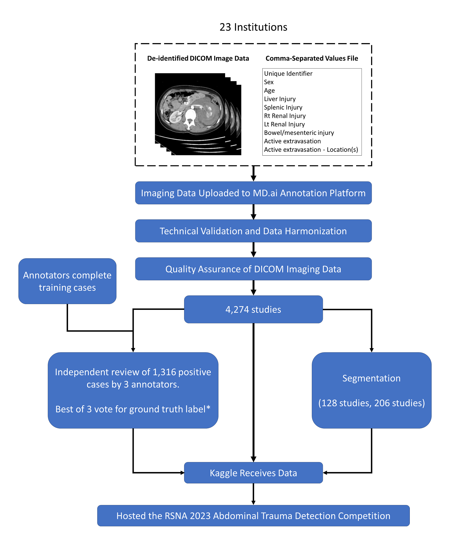
A flowchart of how the RATIC dataset was curated and annotated is shown in Figure 1 with a detailed description provided in Supplementary Materials. In brief, sites provided initial
7
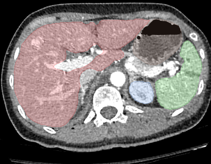
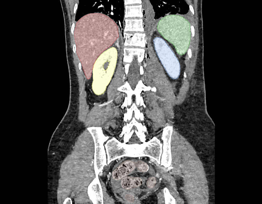
labels for the presence of different traumatic injuries based on clinical reports. Radiologist annotators recruited from the ASER and SAR then annotated solid organ injury grades and locations of bowel/mesenteric injuries and active extravasation. Ground truth labels for the grading of solid organ injuries were established through best of 3 majority voting and collapsed into low (AAST I - III) and high grade (IV and V) injury groups. Image level labels for bowel/mesenteric injuries and active extravasation were based on the consensus of different annotators. Voxel-wise segmentations (Figure 2) were manually corrected after training a nnU- Net (18) on the TotalSegmentator dataset (19), focusing only on the organs being evaluated in the challenge: (1) liver, (2) spleen, (3) left kidney, (4) right kidney, and (5) bowel (representing a combination of esophagus, stomach, duodenum, small bowel, and colon).
Discussion
We curated a large, high-quality dataset of abdominal trauma CTs with contributions from 23 institutions in 14 countries and 6 continents. This represents the largest and most diverse publicly available dataset of abdominal trauma CT scans. This dataset provides annotations relating to injuries of the liver, spleen, kidneys, bowel, and mesentery, as well as active extravasation. This rich dataset has further utility for investigators as other injuries such as hematomas, fractures, and lower thoracic injuries are present within the dataset but were not explicitly annotated due to intent of the challenge to focus on critical findings of the highest clinical importance for trauma patients.
We chose broad inclusion criteria for the contributed CT scans. Our initial survey of potential contributing sites showed great variety in the protocols used for imaging abdominal trauma patients. In fact, some institutions had multiple protocols and selected a protocol based on the severity of the trauma. Aspects that varied across protocols included the parts of the body that were imaged, phases of imaging, and slice thickness. Stringent inclusion criteria that limited contributed scans to a single homogenous protocol (e.g. thin slice, multiphasic CT scans of the
8
abdomen and pelvis) would severely constrain the size and potential generalizability of this dataset. For this reason, we widened the inclusion criteria to facilitate a larger and more diverse dataset that could then be used to train more robust ML models. Biphasic (arterial and portal venous), split bolus, and portal venous phase protocols were considered acceptable.
Participating sites were asked to enrich the dataset with representative injuries given the relatively low prevalence of traumatic abdominal injuries on CT encountered in clinical practice. Despite this request, the number of cases with injuries submitted was lower than the organizing committee had anticipated. Addressing class imbalances in curated datasets is particularly important in improving ML model robustness and reducing bias (20,21). An explicit effort was made by the organizing committee to reduce potential biases in the dataset by considering factors such as sex, age, injuries, and contributing site when assigning scans to the train, public test, and private test datasets.
A challenge we faced in curating this dataset was the dramatic differences in z-axis coverage in the contributed CT scans. For example, some sites imaged from the skull vertex to feet while many sites limited imaging to the abdomen and pelvis. To reduce the size of the dataset and to help ML model training by reducing the search space, we decided to limit scans to the abdomen and pelvis, using an upper bound of the mid-heart and lower bound of the proximal femurs using an automated pipeline (22) and manually reviewed the processed scans.
Similar to prior challenges, we wanted to maximize use of the data and ensure high quality labels while not overburdening annotators. Contributing sites pre-labelled submitted scans with information extracted from the clinical report that allowed annotators to focus on the abnormal scans. We considered a variety of annotation strategies that ranged from study to pixel level annotations. Our past experience with the cervical spine fracture detection challenge (23) showed that the prize-winning models relied on study level annotations and segmentations rather than bounding boxes (24). In addition, recent work has shown that strongly supervised models trained on slice level labels from the RSNA Brain CT Hemorrhage dataset labels (25) do not outperform
9
weakly-supervised models trained on study level labels (26). Pixel level annotations of injuries, including bounding boxes, would be a time consuming task with likely poor reproducibility as abdominal injuries can be quite complex with ill-defined borders. We settled on providing segmentations of the relevant abdominal organ systems to assist with localization and organ labels at the study level. Image level labels were provided for bowel/mesenteric injuries and active extravasation as these injuries can be subtle, present in variable anatomic locations, and present on a limited number of images.
Individual annotators were assigned a single organ system to annotate rather than providing annotations for multiple organ systems on their assigned CT scans. The organizing committee felt this would improve the efficiency of the annotation process and label quality by allowing an annotator to focus on a single task and AAST injury grading scale. The annotators provided granular labels using the AAST grading scale for solid organ injuries. Due to the well documented issues with inter-rater agreement in the grading of solid organ injuries with AAST (9) and to help model training, AAST grades I - III were collapsed into low grade injuries while grades IV and V were considered high grade injuries. This grouping of injury grades still provides more information than a binary label for injury and reflects many clinical practices where grade IV and V injuries are more likely to undergo surgery or endovascular treatment (4-6). Rather than assigning a fixed number of CT scans for annotation, we utilized the crowd sourcing mode on the annotation platform. This allowed annotators to label as many cases as they wanted with the public scoreboard providing motivation.
Each solid organ injury label was annotated independently by 3 radiologists and the final ground truth labels were established by majority. In scenarios where all 3 annotators assigned different gradings (i.e. no injury, low grade, and high grade), a member of the organizing committee adjudicated the case and assigned the final ground truth label. We felt that this approach would improve annotation quality by generating labels with better inter-rater agreement and avoiding the problem of a poor-quality annotation in a single annotation scheme. With an
10
approach that relies on a single annotator per scan, it can be difficult to detect poor quality annotators following completion of the trial of training cases. There are several limitations of this dataset. Ground truth labels for the grading of solid organ injuries were established through best of 3 majority voting. While this represents an improvement over a single annotator, there are inherent issues with AAST grading as a result of inter-rater variability. We recognize the absence of delayed phase imaging as a limitation since it forms part of the AAST imaging criteria for grade II - IV renal injuries in terms of collecting system injuries. Delayed phase imaging was not included as it was not part of the routine protocol for most contributing sites and we were concerned that its inclusion in cases with renal injuries would bias models potentially through spurious associations rather than truly detecting collecting system injuries. Finally, ground truth labels for solid organ injuries, bowel and mesenteric injuries, and active extravasation were made using a web-based annotation platform which is limited when compared to real world clinical practice with access to high resolution monitors, multi-planar thin slice imaging, clinical information, and prior imaging examinations.
In summary, the RATIC dataset represents the largest and most geographically diverse, publicly available expert-annotated dataset of abdominal traumatic injury CT studies. With the release of this dataset, we hope to facilitate research and development in machine learning and abdominal trauma that can lead to improved patient care and outcomes. This dataset is made freely available to all researchers for non-commercial use.
Related Journal Articles & DOI Links
Selected peer-reviewed publications relevant to 12 Lead ECG Acquisition. Click the DOI to access the full paper (may require institutional access).
-
1. Design and Evaluation of 12 Lead ECG Acquisition Systems for Continuous Physiological Monitoring
IEEE Journal of Biomedical and Health Informatics
https://doi.org/10.1109/JBHI.2020.2981234 -
2. Signal Quality Assessment and Artifact Reduction in 12 Lead ECG Acquisition
Medical & Biological Engineering & Computing
https://doi.org/10.1007/s11517-020-02145-6 -
3. Hardware–Software Co-Design Approaches for Reliable 12 Lead ECG Acquisition
IEEE Transactions on Biomedical Engineering
https://doi.org/10.1109/TBME.2019.2895762 -
4. Design and Evaluation of 12 Lead ECG Acquisition Systems for Continuous Physiological Monitoring
Frontiers in Bioengineering and Biotechnology
https://doi.org/10.3389/fbioe.2020.00123 -
5. Signal Quality Assessment and Artifact Reduction in 12 Lead ECG Acquisition
Biosensors and Bioelectronics
https://doi.org/10.1016/j.bios.2021.112345 -
6. Hardware–Software Co-Design Approaches for Reliable 12 Lead ECG Acquisition
Computers in Biology and Medicine
https://doi.org/10.1016/j.compbiomed.2021.104567 -
7. Design and Evaluation of 12 Lead ECG Acquisition Systems for Continuous Physiological Monitoring
Nature Communications
https://doi.org/10.1038/s41467-020-12345-6
Why Choose Us?
Bangalore guidance for robotics, Spectre and autonomous systems projects.
Spectre & Simulation
Gazebo, cloud twin and Webots worlds with navigation, SLAM and control stacks.
Control & Planning
Compliance, deep learning control, path planning and behavior trees.
Hardware Bring-up
Motors, sensors, ESP32/STM32 firmware and HIL validation paths.
Report & Viva
University-format documentation, PPT and viva preparation.
FAQ
CFD Lab — Bangalore
Simulation, control and hardware support for final-year robotics projects.
Stacks
Worlds
Digital Twin
Control
Robots
Offline
Bring-up