Models⋆
Christopher R. Butson1, Scott E. Cooper2, Jaimie M. Henderson3,
Cleveland, Oh
2 Center for Neurological Restoration, Cleveland Clinic Foundation, Cleveland, OH Abstract. Deep brain stimulation (DBS) is an established therapy for the treatment of movement disorders, and has shown promising results for the treatment of a wide range of other neurological disorders. How- ever, little is known about the mechanism of action of DBS or the volume of brain tissue affected by stimulation. We have developed methods that use anatomical and diffusion tensor MRI (DTI) data to predict the vol- ume of tissue activated (VTA) during DBS. We co-register the imaging data with detailed finite element models of the brain and stimulating electrode to enable anatomically and electrically accurate predictions of the spread of stimulation. One critical component of the model is the DTI tensor field that is used to represent the 3-dimensionally anisotropic and inhomogeneous tissue conductivity. With this system we are able to fuse structural and functional information to study a relevant clinical prob- lem: DBS of the subthalamic nucleus for the treatment of Parkinsons disease (PD). Our results show that inclusion of the tensor field in our model caused significant differences in the size and shape of the VTA when compared to a homogeneous, isotropic tissue volume. The magni- tude of these differences was proportional to the stimulation voltage. Our model predictions are validated by comparing spread of predicted activa- tion to observed effects of oculomotor nerve stimulation in a PD patient.

In turn, the 3D tissue electrical properties of the brain play an important role in regulating the spread of neural activation generated by DBS.
Introduction
Deep brain stimulation (DBS) is an effective, proven therapy for the treatment of Parkinson’s Disease (PD), essential tremor and dystonia. The realization that ⋆The authors would like to thank Ashu Chaturvedi, Barbara Wolgamuth, and Christo- pher Maks for their technical assistance on this project. This work was supported by grants from the American Parkinson Disease Association, the Ohio Biomedical Re- search and Technology Transfer Partnership, and the National Institutes of Health
(Ns-50449 & Ns-52042).
R. Larsen, M. Nielsen, and J. Sporring (Eds.): MICCAI 2006, LNCS 4191, pp. 429–437, 2006.
430
C.R. Butson et al. high frequency DBS generates clinical benefits analogous to those achieved by surgical lesioning has transformed the use of functional neurosurgery for the treatment of movement disorders . Thalamic DBS for intractable tremor has virtually replaced ablative lesions of the thalamus . And, DBS of the subthalamic nucleus (STN) or globus pallidus internus has largely replaced pal- lidotomy in the treatment of the cardinal motor features of PD (resting tremor, rigidity, bradykinesia) . In addition, multiple pilot studies have begun to exam- ine the utility of DBS for dystonia, epilepsy, obsessive-compulsive disorder, and depression. However, the clinical successes of DBS are tempered by our limited understanding of the effects of DBS on the nervous system, and the therapeutic mechanisms of DBS remain unknown .
The fundamental purpose of DBS is to modulate neural activity with extracel- lular electric fields, but the technology necessary to accurately predict and visu- alize the neural response to DBS has not been previously available. In turn, the current state-of-the-art in pre-operative targeting strategies and post-operative parameter selection processes do not take into account the influence of the elec- tric field generated by the DBS electrode on the surrounding neural structures.
DBS systems provide thousands of possible stimulation configurations, resulting in great flexibility in customization of therapy for individual patients, but at the expense of time-consuming clinical evaluation to determine therapeutic stimu- lation settings . As a result, DBS patient management is primarily based on trial-and-error as opposed to quantitative understanding. To address these lim- itations, we have developed techniques to predict the volume of tissue activated (VTA) during DBS . We have previously shown that the electrode-tissue inter- face and the encapsulation layer surrounding the electrode can cause substantial differences in the VTA volume. The electrode-tissue interface interacts with the time course of the stimulation waveform such that longer pulse widths are more strongly attenuated . Similarly, high electrode impedances are correlated with greater electrode encapsulation, which also reduces the VTA volume . How- ever, these previous studies were conducted using a homogeneous, isotropic tissue medium which is useful for parameterizing the effects of DBS but does not take into account the complex properties of neural tissue. In this paper we explore the effects of 3-dimensional tissue electrical properties on the VTA generated by DBS. Specifically, tissue conductivity is extracted from diffusion tensor MRI (DTI) data and coregistered with pre-operative and post-operative anatomical MRIs. With this method we are able to create a realistic volume conductor for use in a finite element field model of the brain . Until now there have been few methods available to predict or visualize the effects of DBS on individual patient brains. The innovation of this paper is to predict the volume of tissue activated during stimulation using quantitative forward field models. This type of model is potentially useful to the EEG/MEG community because it provides a forward field solution to brain dynamics that can be used to validate the in- verse models often used for localizing neural activity. The results presented in this paper are the first clinically validated, patient-specific models of DBS for predicting neural activation.
Methods
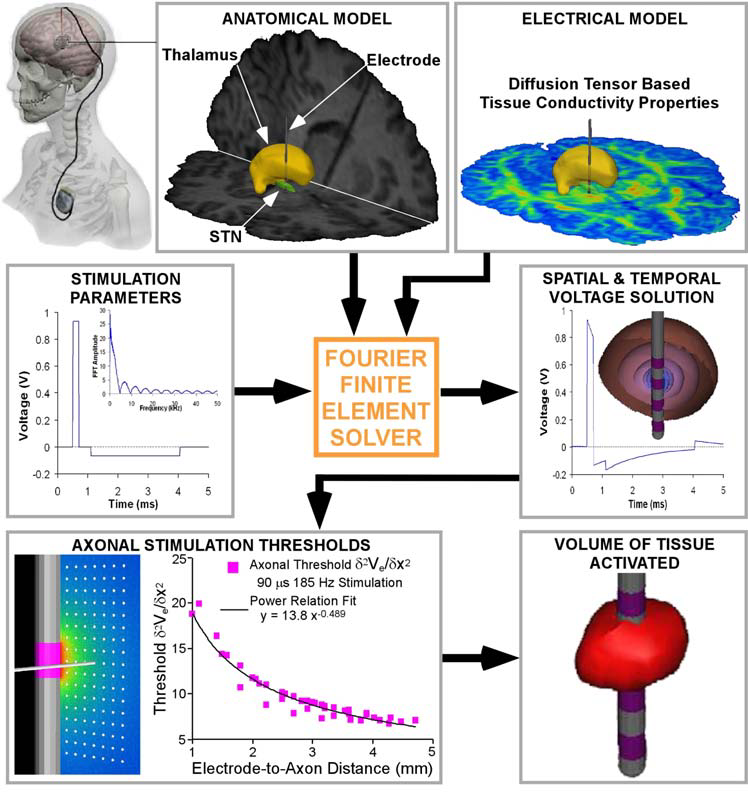
The DBS system consists of a quadripolar electrode lead, an implantable pulse generator (IPG) and an extension wire connecting the two (Figure 1). The elec- trode lead is implanted during stereotactic surgery; the work presented in this paper focuses on STN DBS. Each electrode contact is 1.27 mm diameter and 1.5 mm height, and electrodes are separated by either a 1.5 mm (3387 model) or 0.5 mm (3389 model) insulation gap. The DBS system can be operated in multi-polar or monopolar configurations with any combination of anodes and cathodes; during monopolar stimulation the return electrode is the IPG case.
Typical settings for monopolar STN DBS are -3 V, 90 μsec pulse width and 130 Hz. For therapeutic stimulation settings, symptomatic relief of PD symptoms is normally achieved within a few seconds.
We have developed techniques to predict the spread of activation during DBS by combining several imaging methods into a single coherent framework (Figure 1). First, a pre-operative T1 MRI (collected on a Siemans 1.5T scanner) was segmented using SNT software (Surgical Navigation Technologies, a division of Medtronic, Minneapolis, MN, USA) based on the non-linear warping techniques of to produce closed surfaces for several basal ganglia nuclei: STN, thalamus, globus pallidus, putamen, caudate and accumbens (only STN and thalamus are shown in the figures). Second, a post-operative T1 MRI was used to determine the position of the electrode. Electrode localization was performed by isosurfac- ing progressively smaller values of the electrode artifact until the surface con- verged onto a volume containing the 4 electrode contacts. The pre-operative and post-operative MR volumes were then co-registered with fractional anisotropy values from a DTI atlas brain using Analyze 6.0 (AnalyzeDirect, Lenexa, KS, USA). The DTI atlas brain is explained in detail in . Briefly, images were acquired on a 1.5-T Philips Gyroscan MR using single-shot echo-planar imaging sequences. The imaging matrix was 112 x 112 with a field of view of 246 x 246 mm. Transverse sections of 2.2 mm thickness were acquired for a total of 55 slices. Diffusion weighting was encoded along 30 independent orientations, and the b value was 700mm2/sec. Imaging was repeated six times to enhance the signal to noise ratio. The six independent elements of the 3 x 3 diffusion tensor were calculated for each pixel by using a multivariate linear fitting method.
Accurate representation of the stimulating electrodes and the surrounding elec- tric field is made possible with two techniques: multiresolution meshes and dy- namic interpolation. Finite element methods are used to model geometric features and boundary conditions for which analytical solutions do not exist. A finite ele- ment model of the DBS electrode and surrounding tissue medium was constructed using FEMLAB 3.1 (Comsol Inc., Burlington, MA, USA). Multi-resolution mes- hes allow node density to be highest where the electric field is changing the fastest.
The second technique, dynamic interpolation, allows the electrode to be easily moved to arbitrary positions and orientations within the brain volume without repeating the computationally costly and time-consuming step of creating a new mesh. With these methods, DTI tensors are dynamically interpolated onto the fi- nite element mesh, an operation that takes 10-15 seconds for approximately
432
C.R. Butson et al. Fig. 1. Methods. The DBS system is implanted as shown. Pre-operative, post-operative and DTI MRIs are coregistered, the stimulation waveform is applied to the electrode, and the system is solved using the Fourier FEM solver. The resulting time- and space- dependent voltages are used to calculated activating function thresholds for fibers of passage, and the VTA is determined using these thresholds.
215,000 nodes and 1.9e6 DTI tensors on an 8-processor SGI Prism with 32 GB RAM. Lastly, boundary conditions for the stimulation parameters at each elec- trode contact are dynamically interpolated onto the brain mesh.
The conductivity tensor is directly solved for at each voxel using a simple linear transform : σ = (σe/de)D, where σe is the effective extracellular con- ductivity, de is the effective extracellular diffusivity and D is the diffusion tensor.
The ratio of (σe/de) has been determined from experimental and empirical data to be ~0.7 (S - s)/mm2 . This value is then scaled to match the electrode impedance recorded from the IPG of the given patient.
Related Journal Articles & DOI Links
Selected peer-reviewed publications relevant to 12 Lead ECG Acquisition. Click the DOI to access the full paper (may require institutional access).
-
1. Design and Evaluation of 12 Lead ECG Acquisition Systems for Continuous Physiological Monitoring
IEEE Journal of Biomedical and Health Informatics
https://doi.org/10.1109/JBHI.2020.2981234 -
2. Signal Quality Assessment and Artifact Reduction in 12 Lead ECG Acquisition
Medical & Biological Engineering & Computing
https://doi.org/10.1007/s11517-020-02145-6 -
3. Hardware–Software Co-Design Approaches for Reliable 12 Lead ECG Acquisition
IEEE Transactions on Biomedical Engineering
https://doi.org/10.1109/TBME.2019.2895762 -
4. Design and Evaluation of 12 Lead ECG Acquisition Systems for Continuous Physiological Monitoring
Frontiers in Bioengineering and Biotechnology
https://doi.org/10.3389/fbioe.2020.00123 -
5. Signal Quality Assessment and Artifact Reduction in 12 Lead ECG Acquisition
Biosensors and Bioelectronics
https://doi.org/10.1016/j.bios.2021.112345 -
6. Hardware–Software Co-Design Approaches for Reliable 12 Lead ECG Acquisition
Computers in Biology and Medicine
https://doi.org/10.1016/j.compbiomed.2021.104567 -
7. Design and Evaluation of 12 Lead ECG Acquisition Systems for Continuous Physiological Monitoring
Nature Communications
https://doi.org/10.1038/s41467-020-12345-6
Why Choose Us?
Bangalore guidance for robotics, Spectre and autonomous systems projects.
Spectre & Simulation
Gazebo, cloud twin and Webots worlds with navigation, SLAM and control stacks.
Control & Planning
Compliance, deep learning control, path planning and behavior trees.
Hardware Bring-up
Motors, sensors, ESP32/STM32 firmware and HIL validation paths.
Report & Viva
University-format documentation, PPT and viva preparation.
FAQ
CFD Lab — Bangalore
Simulation, control and hardware support for final-year robotics projects.
Stacks
Worlds
Digital Twin
Control
Robots
Offline
Bring-up