This version has been peer-reviewed and accepted for publication by Elsevier Towards a better understanding of abdominal wall biomechanics: in vivo relationship between dynamic intra-abdominal pressure and
1,4
1Univ Gustave Eiffel, Aix-Marseille Univ, LBA, F-13016 Marseille, France 2Aix Marseille Univ, CNRS, CRMBM UMR 7339, Marseille France 3Assistance Publique-Hopitaux de Marseille, Hôpital Universitaire Timone, CEMEREM, Marseille, France
Victoria Joppin
Laboratoire de Biomécanique Appliquée, UMRT24 Université Gustave Eiffel - Aix Marseille Université Faculté des Sciences Médicales et Paramédicales - Secteur Nord 51 Boulevard Pierre Dramard, F-13016 Marseille, France
Highlights
• Abdominal wall pattern by coupling magnetic resonance image and pressure sensor • Correlation study of muscle motion, visceral area and intra-abdominal pressure change • Lateral muscles were correlated with pressure for active exercise (cough, Valsalva) • Rectus muscles were correlated with pressure for passive exercise (breathing) • Highlights the impact of individual mechanical properties on physiological response
Arxiv:2508.12827V2 [Physics.Med-Ph] 30 Jan 2026
This version has been peer-reviewed and accepted for publication by Elsevier
Abstract
Background: In vivo mechanical behaviour of the abdominal wall has been poorly characterised and important details are missing regarding the occurrence and post-operative recurrence rate of hernias which can be as high as 30%. This study aimed to assess the correlation between abdominal wall displacement and intra-abdominal pressure, as well as abdominal compliance.
Methods: Eighteen healthy participants performed audio-guided passive (breathing) and active (coughing, Val- salva maneuver) exercises. Axial dynamic changes of abdominal muscles and visceral area were measured using MRI, and intra-abdominal pressure with ingested pressure sensor.
Findings: Correlations between abdominal wall displacement and intra-abdominal pressure were specific to participant, exercise, and varying between rectus abdominis and lateral muscles. Strong correlations were found between rectus abdominis displacement and intra-abdominal pressure during breathing (r = 0.92 ± 0.06), as well as lateral muscles displacement with intra-abdominal pressure during coughing and Valsalva maneuver (r = −0.98 ± 0.03 and −0.94 ± 0.05 respectively). The abdominal pseudo-compliance varied greatly among participants during muscular contraction, the coefficient of variation reaching up to 70%.
Interpretation: The combination of intra-abdominal pressure and dynamic MRI measurements enables the identification of participant-specific behaviour pattern. Intra-abdominal pressure and abdominal wall dynamic undergo consistent and predictable interactions. However, this relationship is subject-specific and may not be extrapolated to other individuals. Therefore, both intra-abdominal pressure and abdominal wall motion must be measured in the same participant in order to accurately characterise the abdominal wall behaviour. These results are of great importance for mesh design, surgical decision-making, and personalised healthcare.
2
This version has been peer-reviewed and accepted for publication by Elsevier
Keywords
Abdominal wall muscles; Biomechanics; Dynamic magnetic resonance imaging; Intra-abdominal pressure; Me-
Abbreviations
IAP: intra-abdominal pressure; LM: lateral muscles; RA: rectus abdominis; MRI: magnetic resonance imaging; p: p-value; r: Pearson correlation coefficient; CV: coefficient of variation; Cab: Pseudo abdominal compliance
3
This version has been peer-reviewed and accepted for publication by Elsevier
Introduction
The abdominal wall is a multi-layered structure crucial for organ protection, upper body movement and intra- abdominal pressure (IAP) control. However, our understanding of the relationship between IAP and abdominal wall motion is limited, largely due to inadequate investigative methods. Enhanced knowledge of abdominal wall mechanical behaviour could provide valuable insights into physiological and pathological phenomena, such as hernia development. Hernias, characterised by the protrusion of abdominal contents through defects in the musculo-aponeurotic structure, can cause pain and diminish quality of life . Despite advances in surgical treatment, hernia recurrence remains high i.e. 30% [2, 3], likely due to a conflict between the increased IAP leading to an increased abdominal wall loading and the ability of the abdominal wall to cope with this load by moving and deforming.
Abdominal wall displacements and strains have been assessed in vivo mostly externally by image correlation. These studies quantified abdominal motion during exercises [4, 5] and showed that strain depends on anatomical zones [6, 7]. However, to fully understand hernia pathophysiology, a detailed examination of the different ab- dominal wall layers is necessary. Recent research using 2D dynamic MRI has reported specific abdominal muscle displacements . While these studies provide valuable data, integrating IAP is critical to fully understand the relationship between abdominal wall motion and pressure, which is essential for understanding both normal and pathological conditions such as hernias.
Most studies on IAP have focused on intraperitoneal hypertension and abdominal compartment syndrome , with fewer investigations into its role in hernia formation, although it has been recognised as a potentially important factor . IAP changes can modulate external abdominal wall surface strain, internal muscular deformations and abdominal volume. Several studies using laparoscopic peritoneal gas insufflation in post- mortem conditions , anesthetised patients and rabbits have confirmed the anisotropic behaviour of the abdominal wall, with greater curvature radius and shear modulus in the longitudinal direction.
Song et al. showed a near linear correlation between antero-posterior displacement of the abdominal wall and increasing IAP . In post mortem human species, Konerding et al. observed a linear correlation between IAP and transverse forces on the linea alba . In vivo studies also showed that IAP was correlated with electrical muscle activity [15, 16].
Abdominal compliance, defined as the ratio of intra-abdominal volume change to IAP change, reflects the abdom- inal wall’s ability to distend in response to pressure . Abdominal compliance has been mainly investigated in vivo during laparoscopic peritoneal gas insufflation in porcine models [18, 19] and humans [18, 20, 21, 22].
In a porcine model, Vlot et al. reported a 93% abdominal volume increase when pressure rose from 5 to 10 mmHg. Using CT and surface image correlation, Becker et al. reported an exponential relationship between in subcutaneous fat thickness and compliance. In healthy humans, body position (upright or supine) and abdominal binders were shown to influence compliance . However, compliance during voluntary muscle activation has yet to be studied.
Despite these advances, gaps remain in our understanding of in vivo real-time interactions between IAP and abdominal wall response. Current clinical practices measure IAP invasively via the bladder, but ingestible sensors offer a safer, non-invasive alternative [24, 25].
Surface imaging techniques are limited to external observations, while ultrasound and elastography only provide localised data. CT scanning, though in-depth, involves radiation. In this context, MRI stands out as a non-invasive, non-radiating alternative that extends the in-depth study of the various muscle groups in the abdominal wall.
To our knowledge, this is the first study to dynamically assess the in vivo relationship between IAP and MRI-based metrics of abdominal wall movement and deformation. Through the combination of dynamic MRI and ingestible pressure sensor measurements in healthy human participants, this study aims to enhance our understanding of the mechanical behaviour of the abdominal wall during various physiological activities that could contribute to hernia development.
Participants
Eighteen healthy participants (8 females) volunteered to take part in the study after providing their informed written consent. The average age and BMI were 29.3 ± 8.1 years old and 22.6 ± 2.5 kg/m2 respectively.
4
This version has been peer-reviewed and accepted for publication by Elsevier This study was approved by the French ethics committee (IDRCB: 2019-A00806–51) and was conducted ac- cording to national legislation related to interventional research and the Declaration of Helsinki.
Protocol
Participants were positioned supine in the MRI scanner (MAGNETOM Verio, Siemens Healthineers, Erlangen, Germany) with integrated and flexible body coils on the abdomen. Dynamic (2D+t) MRI captured axial changes every 182 ms during three audio-guided exercises, as previously described . Breathing (inhale-exhale deeply during 10 s), coughing and the Valsalva maneuver (forceful expiration against closed nostrils and mouth during 8 s), chosen as representative of daily activities and clinical exam , were performed after training.
Breathing was considered passive while coughing and the Valsalva maneuver were active exercises involving abdominal contraction . In a separate session, participants repeated the same exercises after swallowing a SmartPillTM capsule (Medtronic, Minneapolis, MN, USA) to measure IAP (± 3.6 mmHg precision) with a 500 ms sampling time.
Data Processing
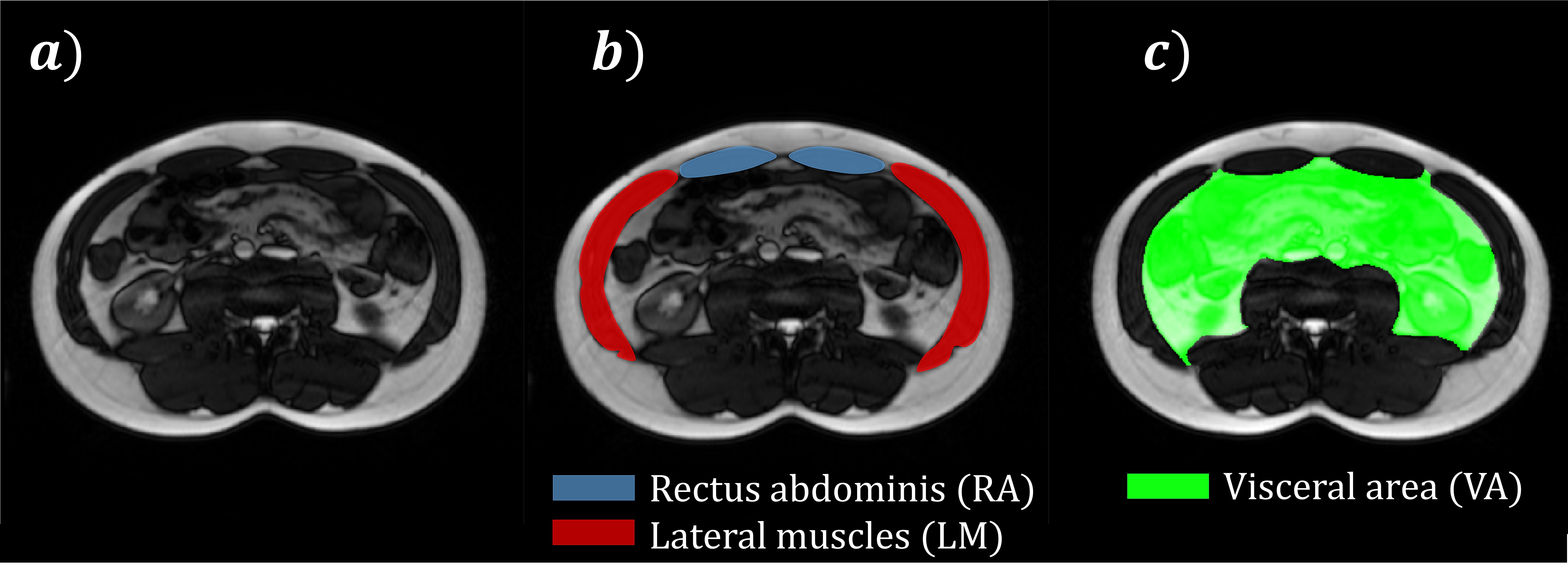
As illustrated in Figure 1-a, muscles of interest were manually delineated in MR images using FSLeyes , then a semi-automatic segmentation algorithm was used to obtain the segmentation masks at each timestep . Segmentation masks included the rectus abdominis (RA) and lateral muscles (LM, including the transverse abdominis, internal and external obliques), as represented in Figure 1-b.
Figure 1: Typical 2D axial abdominal MRI slice without annotation a)
Segmentation Masks Are Indicated In B) And C)
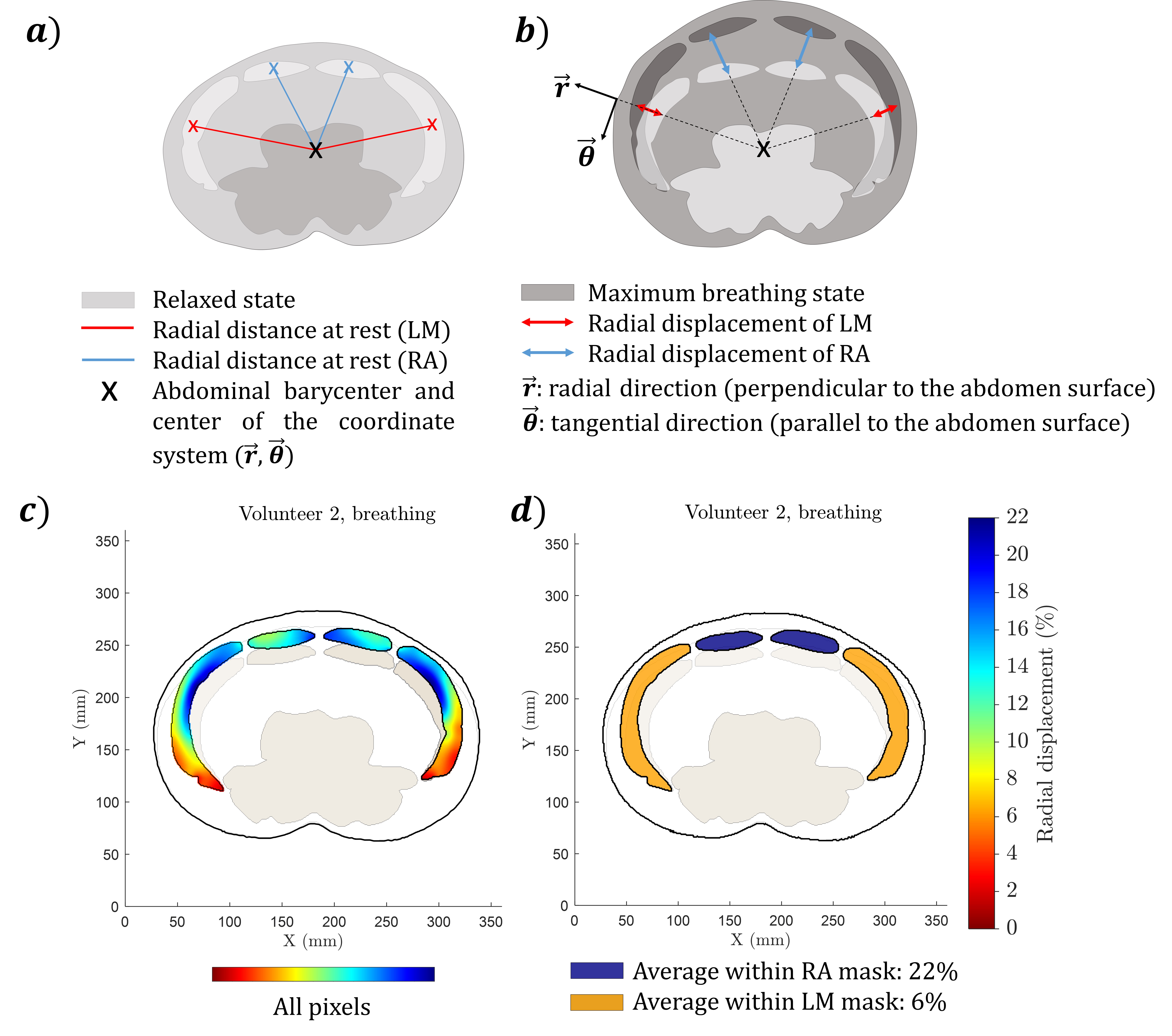
Radial displacements (Figure 2-b) were computed for each pixel of the segmented muscles (Figure 2-c), then averaged across the LM and RA muscles (Figure 2-d).
5
This version has been peer-reviewed and accepted for publication by Elsevier
B) Radial Displacement Scheme
c) Radial displacement of each pixel of the segmentation masks d) Radial displacement averaged among LM and RA segmentation masks The visceral area (Figure 1-c), defined as the region delimited by the internal surface of the abdominal mus- cles and dorsal structures (quadratus lumborum, psoas major, erector spinae, vertebral disc and body), was computed.
Radial displacement of LM and RA muscles and visceral area metrics were normalised to their resting values, defined as the endpoint of a calm exhalation without visible contraction, ensuring no variation at rest. For standardisation between participants, these metrics were expressed relative to the resting state: the visceral area was divided by its resting value, and the radial displacements were divided by the radial distance between the barycenter of each muscle group (LM and RA) at resting state and the abdominal barycenter (Figure 2-a).
The barycenter of a muscle is defined as the mean position of all pixels that constitute its segmentation mask. Meanwhile, the abdominal barycenter is the average location of all pixels composing the LM segmentation masks.
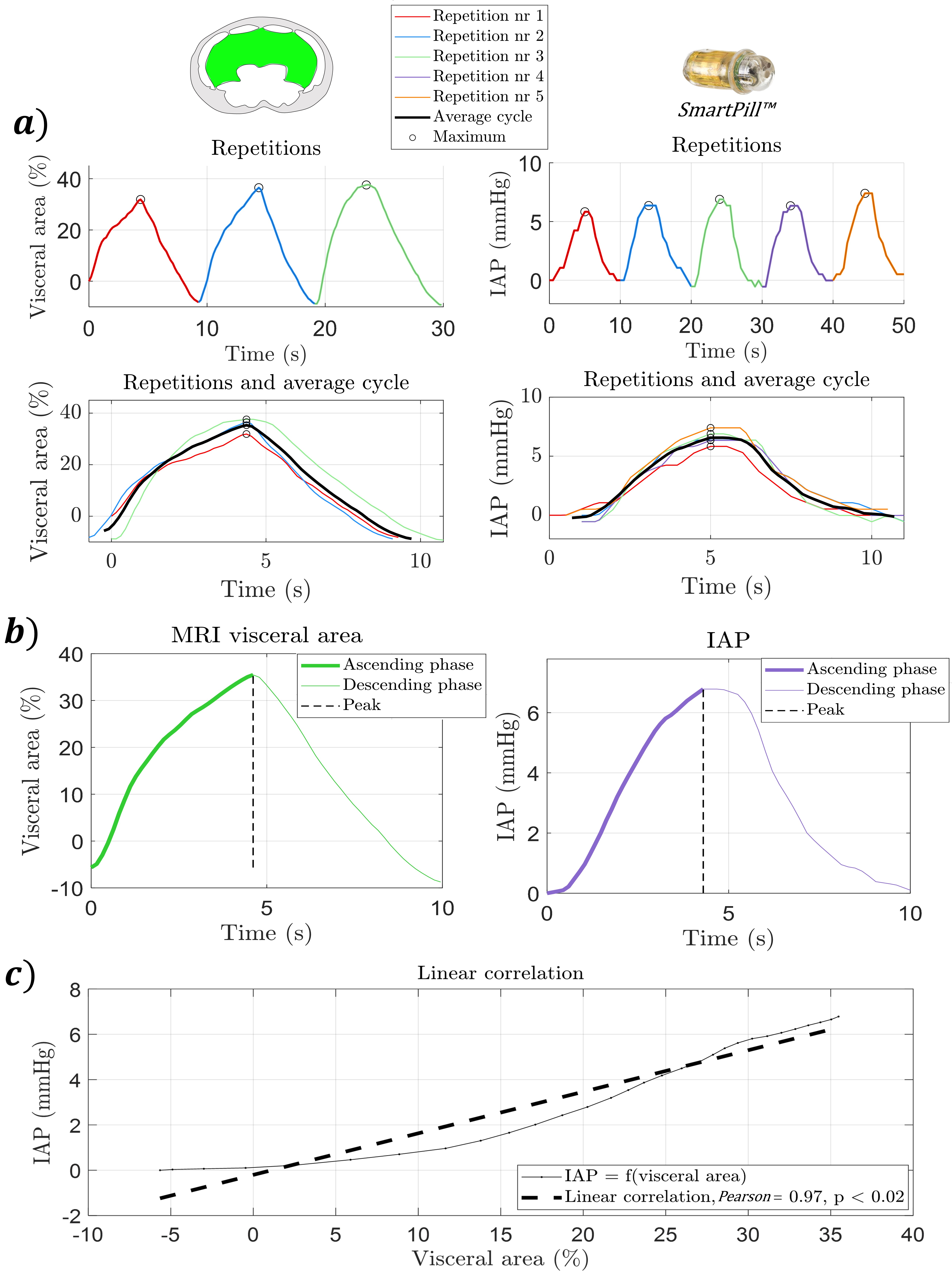
As illustrated in Figure 3-a for each exercise, specific timepoints, i.e. onset and peak, were manually identified so as to delimit the different repetitions using MATLAB. Then, all repetitions were linearly oversampled to n points, corresponding to the longest repetition across participants. This equates to n = 60, 12 and 54 points for breathing, coughing and the Valsalva maneuver, respectively, due to varying exercise durations. This approach was chosen to achieve a slight upsampling of the shorter repetitions, while avoiding the loss of information associated with downsampling the longer ones. For each participant, the resulting repetitions of n points were averaged to generate a representative average cycle of n points of each exercise (Figure 3-b). As illustrated in Figure 3-a, the maximum values of the each set of repetitions were aligned for signal averaging in order to prevent the peak from being smoothed out.
6
This version has been peer-reviewed and accepted for publication by Elsevier Figure 3: Example of selected timepoints and averaging repetitions during breathing a) Repetitions delimitation (left: visceral area, right: intra-abdominal pressure (IAP)) b) Average cycle (left: visceral area, right: IAP) c) Linear correlation between the average cycles of IAP and visceral area during ascending phase, i.e. from the
Beginning Until The Iap Reaches Its Maximum Value
The IAP values were exported using the SmartPillTM 3.1 software with the temperature-compensated mode. The IAP repetitions were oversampled to reach n points by linear interpolation and averaged similarly to the MRI data. This process standardised the cycle lengths across repetitions, allowing for the correlation of MRI-based abdominal data and IAP values, despite non-simultaneous measurements. This approach enabled consistent comparison between abdominal motion and IAP across different participants and exercises.
2.4
Correlation intra-abdominal pressure - abdominal wall motion This study investigates the relationship between intra-abdominal pressure (IAP) and MRI-based metrics of abdominal wall movement and deformation. Pearson correlation coefficients r were calculated for each partici-
7
This version has been peer-reviewed and accepted for publication by Elsevier pant and exercise to evaluate the direction (positive or negative) and strength of correlations between IAP and visceral area, as well as IAP and radial displacement of LM and RA. These correlations were assessed during the ascending phase of each exercise (until maximum or plateau for the Valsalva, as illustrated in Figure 3-c), with statistical significance considered for p-value P ≤0.05 and r ≥0.468, critical threshold of an acceptable correlation strength .
Abdominal pseudo-compliance was computed as the ratio between the maximum changes in visceral area ∆maxV A (value before normalisation, expressed in cm2) and intra-abdominal pressure ∆maxIAP:
Mmhg )
The maximum change was defined as the difference between the beginning and the end of the ascending phase (Figure 3-b). This metric differs from the conventional compliance using volume given that it is applied to 2D MRI slices.
Results were presented as average across all participants (n = 18) ± standard deviation σ, with their [mini- mum/maximum] range and coefficient of variation CV, defined as the standard deviation divided by the absolute r value: CV = σ/|r| (%).
The correlation between IAP and MRI measurements was further analysed in relation to morphologic param- eters, including BMI (kg/m2), waist circumference (mm), radial distance at rest of LM and RA (mm) and muscle mass ratio (%), defined as the area occupied by abdominal muscles divided by the whole abdomen area (delimited by the external surface of the abdomen). The difference according to the sex assigned at birth (female or male) was assessed using a two-sided Wilcoxon-Mann-Whitney test, with a P ≤0.05 considered statistically significant.
3.1
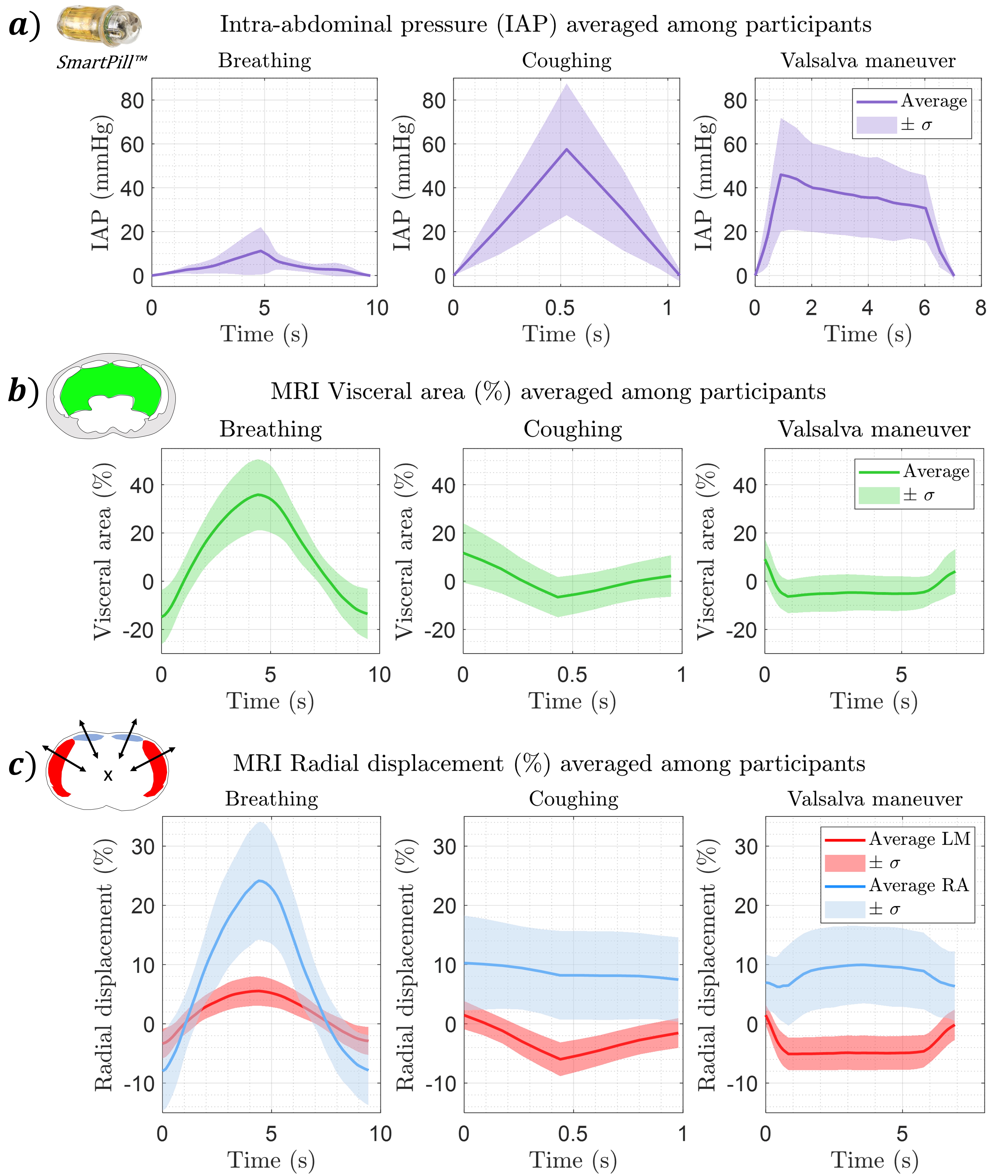
Intra-abdominal pressure and MRI time-dependent changes Temporal changes of IAP, visceral area and radial displacement of LM and RA during the different exercises are displayed in Figure 4, with maximum changes summarised in Table 1. Radial displacement and visceral area were analysed for 17 participants as one MRI dataset was excluded due to poor quality.
8
This version has been peer-reviewed and accepted for publication by Elsevier Figure 4: Temporal changes for different exercises of
C) Radial Displacement Of Lm And Ra (%)
Results are presented as mean values ± one standard deviation σ During breathing, IAP, visceral area and radial displacement of LM and RA muscles followed a bell shape pattern with ascending and descending phases. Breathing induced a large visceral area increase and an outward/positive radial displacement.
Coughing also exhibited these two phases, but visceral area and LM displacement initially decreased during contraction, contrasting with IAP’s initial rise. Radial displacement of RA was limited (−2.63 ± 6.38%) compared to LM (−7.32 ± 2.86%), with notable inter-participant variability, RA being pulled in or out according to the participant’s coughing execution. The positive RA displacement at the onset was related to a short intake of air before coughing.
For the Valsalva maneuver, three phases where observed: IAP and RA displacement increased initially, while LM displacement and visceral area decreased. Variability in RA displacement was higher than LM, highlighting again differences in execution among participants (standard deviation of 6.15% for RA compared to 2.99% for
Lm).
Activities with muscular activation led to a visceral area decrease accompanied by an inward, negative displace- ment of LM and a higher ∆maxIAP. IAP during coughing was 11 mmHg higher than during the Valsalva maneuver. For the Valsalva maneuver, a significant correlation between ∆maxIAP and the muscle mass ratio was found (r = 0.71, P ≤0.05), with males showing higher IAP compared to females (59.14 and 29.49 mmHg, respectively).
9
This version has been peer-reviewed and accepted for publication by Elsevier
−1.14 ± 6.15
Table 1: Maximum changes in intra-abdominal pressure ∆maxIAP, visceral area ∆maxV A and radial
Displacement Of Lm And Ra For Different Exercises
Results are presented as mean values ± one standard deviation σ
3.2
Relationship between intra-abdominal pressure and visceral area The relationship between IAP and visceral area changes was assessed in order to provide information related to the dynamic behaviour of the abdominal wall on a global scale, i.e. without distinction between LM and RA muscles. The mean r values are presented in Table 2.
7.61
Table 2: Pearson correlation coefficients (r) for the correlations between intra-abdominal pressure (IAP) and visceral area, range [min/max] and coefficient of variation (CV) Results are presented for the different exercises as mean values ± one standard deviation σ The correlation between IAP and visceral area changes was significant (P ≤0.05 and |r| ≥0.468) and very subject-specific for each participant and each exercise. For coughing and the Valsalva maneuver, r values were negative indicating that IAP was increasing while the visceral area was decreasing. The correlation was strong, with an absolute mean r value larger than 0.9 for the whole set of exercises.
Figure 5 illustrates the maximum changes of intra-abdominal pressure ∆maxIAP and visceral area ∆maxV A for all the participants and the three exercises. Both variables were normalised by their highest values among the 18 participants to be scaled from 0 to 1. The largest IAP change did not always correspond to the largest visceral area change. For example, participant 7 had large area changes with moderate IAP during breathing, whereas participant 5 showed the opposite during the Valsalva maneuver. No significant correlation was found between participants between maximum changes in IAP and visceral area, indicating that their relationship is participant-specific and cannot be generalised to the whole cohort.
Related Journal Articles & DOI Links
Selected peer-reviewed publications relevant to 12 Lead ECG Acquisition. Click the DOI to access the full paper (may require institutional access).
-
1. Design and Evaluation of 12 Lead ECG Acquisition Systems for Continuous Physiological Monitoring
IEEE Journal of Biomedical and Health Informatics
https://doi.org/10.1109/JBHI.2020.2981234 -
2. Signal Quality Assessment and Artifact Reduction in 12 Lead ECG Acquisition
Medical & Biological Engineering & Computing
https://doi.org/10.1007/s11517-020-02145-6 -
3. Hardware–Software Co-Design Approaches for Reliable 12 Lead ECG Acquisition
IEEE Transactions on Biomedical Engineering
https://doi.org/10.1109/TBME.2019.2895762 -
4. Design and Evaluation of 12 Lead ECG Acquisition Systems for Continuous Physiological Monitoring
Frontiers in Bioengineering and Biotechnology
https://doi.org/10.3389/fbioe.2020.00123 -
5. Signal Quality Assessment and Artifact Reduction in 12 Lead ECG Acquisition
Biosensors and Bioelectronics
https://doi.org/10.1016/j.bios.2021.112345 -
6. Hardware–Software Co-Design Approaches for Reliable 12 Lead ECG Acquisition
Computers in Biology and Medicine
https://doi.org/10.1016/j.compbiomed.2021.104567 -
7. Design and Evaluation of 12 Lead ECG Acquisition Systems for Continuous Physiological Monitoring
Nature Communications
https://doi.org/10.1038/s41467-020-12345-6
Why Choose Us?
Bangalore guidance for robotics, Spectre and autonomous systems projects.
Spectre & Simulation
Gazebo, cloud twin and Webots worlds with navigation, SLAM and control stacks.
Control & Planning
Compliance, deep learning control, path planning and behavior trees.
Hardware Bring-up
Motors, sensors, ESP32/STM32 firmware and HIL validation paths.
Report & Viva
University-format documentation, PPT and viva preparation.
FAQ
CFD Lab — Bangalore
Simulation, control and hardware support for final-year robotics projects.
Stacks
Worlds
Digital Twin
Control
Robots
Offline
Bring-up