Mustafa Şen1,2
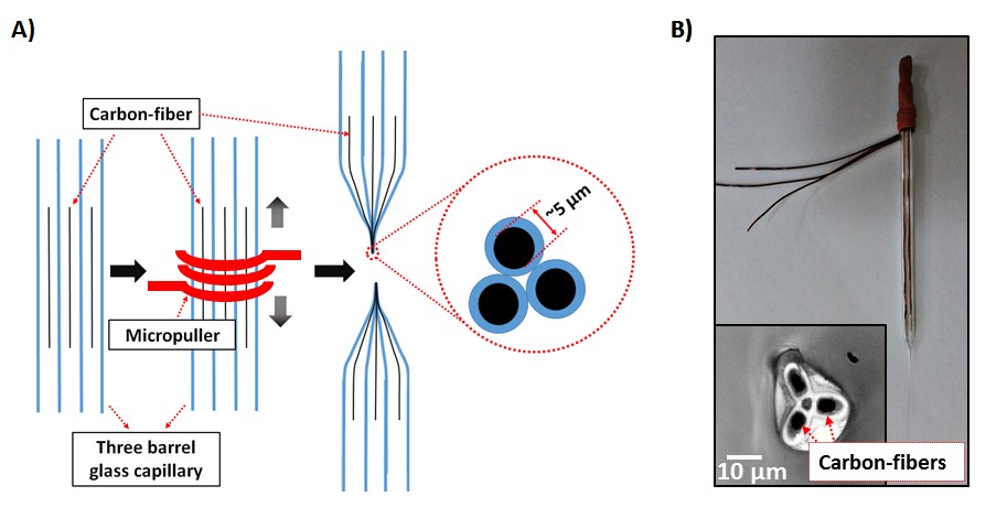
Here, we report a facile method for electro-addressable functionalization of a probe comprising of closely spaced three individually addressable carbon fiber electrodes for detection of nucleic acids. First, a multi electrode array probe comprising three adjacent carbon fiber electrodes was fabricated through pulling a three-barrel glass capillary with a single carbon fiber in each barrel using a micropuller.
Second, electropolymerization based doping was used for electro-addressable functionalization of the multi-electrode array probe. To demonstrate that the current strategy works, anti-miR-34a was electrografted on only one of three electrodes by electropolymerization of pyrrole on the specific electrode. A second electrode was coated only with polypyrrole (PPy) and the third was left unmodified.
Electrochemical impedance spectroscopy (EIS) was used for analysis and the results clearly showed charge transfer resistance of the PPy + anti-miR-34a modified electrode increased significantly after hybridization, while charge transfer resistance of the other two electrodes remained almost constant. The results demonstrate that the present strategy has great potential for constructing multiplex nucleic acid micro/nano biosensors for local and in situ detection of multiple nucleic acid molecules such as miRNAs at a time.
The Use Of Microelectrodes In Construction Of
microbiosensors has many advantages such as the ability to achieve high temporal and spatial resolution, local detection and detection in small volumes1. Increasing the number of electrodes enables detection of multiple analytes or simultaneous and high-throughput detection of same molecules at multiple points. Even though large number of individually addressable electrodes can be effectively assembled in a small area2-5, electrochemical detection methods have a relatively limited ability to detect multiple analytes, particularly when compared to microdialysis probes and other analytic techniques based on chromatography or electrophoretic separation 6. There are several reports where multi-electrode arrays were assembled on a substrate to form a biosensor chip to perform multiple analysis. For example, Zhu et al. used a multi-point addressable electrochemical device where row and column electrodes were orthogonally arranged to form a 4 x 4 array for amperometric detection of DNA hybridization.7 Because of the large distance between electrodes, they were able to modify electrodes with different probes using a pipette. The device had a detection limit of 30 nM. In another study, Erdem et al. used a multi-channel screen printed array electrode for label-free voltammetric detection of microRNAs.8 Electrode array comprising 16 relatively large carbon electrodes enabled 16 consecutive analysis with a detection limit of 4.3 pmole in 3 µL sample.
The treatment of the electrodes with different samples was also done by pipetting due to large distance between the electrodes. A microelectrode array comprising closely spaced electrodes with different labels enables simultaneous and local detection of different analytes in small volumes, but labeling closely spaced electrodes with different bio- recognition molecules is technically challenging as pipetting is not an option. In a recent study, Ozsoz at al. used polypyrrole (PPy) for the doping of probe miRNA on a large pencil graphite electrode to detect miR-219. PPy is one of the most widely used conducting polymer for biosensor design due to its biocompatibility, high electrical conductivity, wide pH range, fast electrochemical reaction, high surface energy and low cost. Basically, in the study, the miRNA probe was mixed with the pyrrole solution and by electropolymerization of pyrrole, the miRNA probes were electrografted on the electrode surface. The biosensor showed good selectivity and had a detection limit of 0.17 nM. Since pyrrole is electropolymerized only on the electrode through which the electropolymerization potential is applied, we used it for electro-addressable functionalization of a closely spaced multi carbon fiber electrode array micro-probe with a pitch of less than 5 µm for the detection of miR-34a. Carbon fiber based microelectrodes are commonly used for local and highly sensitive detection of neurotransmitters such as dopamine or recording of neuronal action potentials known
Of
neurochemical activity of brain 10-13. In addition to being used in such measurements, CFEs have also been used for detection of biologically important molecules with great sensitivity and selectivity by simply modifying the surface of the electrode with a bio-recognition molecule 14-17. To the best our knowledge this is the first study to use a probe-type microelectrode array for nucleic acid detection. The probe was used in self-referencing mode18, for detection of miR- 34a, where one of three electrodes was modified with anti- miRNA34a using PPy. A second electrode was coated with PPy and the third was left unmodified. The results clearly demonstrate that electropolymerization based doping works for electro-addressable functionalization of a micro-electrode array probe and has a great potential for constructing micro- biosensors for detection of multiple miRNAs at a time For fabrication of the multi-electrode array probe, three copper wires, each connected to a single carbon fiber, were prepared. Each wire was prepared as follows; a single carbon fiber with a diameter of 5-6 µm was fixed to an insulated copper wire. To make the connection, one end of the copper wire was dipped into Ag paste that was then used for attaching a single carbon fiber. The Ag paste was cured at 180 °C for 30 min to make a rigid connection between the Using electropolymerization based doping for electro-addressable functionalization of a multi-
2
carbon fiber and the copper wire. Three copper wires, each fixed with a carbon fiber, were individually inserted into a three-barrel capillary. To fabricate a multi-electrode array probe with a thin and parallel run to the very end of the tip, the three-barrel capillary was pulled (PG10165-4, World Precision Instrument, USA) with a micropuller (PC-10, Narishige, Japan) using the one-stage pull option to seal the carbon fibers in glass (Figure 1A)1,19-21. The heating parameter that yielded a multi-electrode array probe with the desired characteristics was 68 °C. In the following step, the excessive unsealed carbon fibers at the tip of the probe were removed with a scissor. To fill any possible gap between the carbon-fibers and the sealed glass, the tip of the probe was dipped into resin which was then cured at 80 °C for 40 min.
To fabricate micro-disk electrodes, the tip of the probe was ground using a microgrinder (EG-401, Narishige, Japan) (Figure 1B).
The Electrochemical Behavior Of The Carbon Fiber
electrodes was analyzed to assess the usability of the probe. First, cyclic voltammograms (CVs) of each electrode were obtained by sweeping the potential from 0 to +0.6 V vs.
A
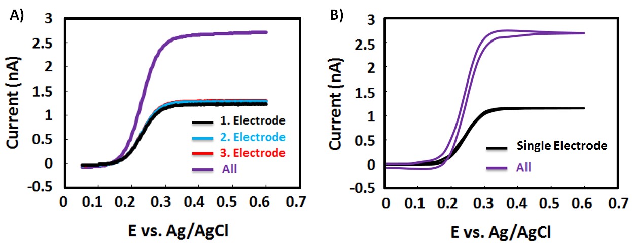
simultaneous CV of all electrodes was obtained under the same conditions. All three electrodes showed similar behavior; the maximum current was measured to be ~1.24 nA (Figure 2A), quite close to the simulated current value of ~1.16 nA (Figure 2B) found using COMSOL Multiphysics.
We believe the difference between experimental and simulated values is normal considering the likely non-ideal surface conditions of the tips of the probe. When all three electrodes were connected, the maximum current measured was ~2.71 nA (Figure 2A), approximately 2.2 times of the current obtained from a single electrode and quite close to the simulated value of ~2.74 nA (Figure 2B). These results clearly indicate that all electrodes are working properly and individually addressable.
After confirming the integrity of the electrodes, the multi-electrode array probe was modified for miR-34a detection using electropolymerization based doping (Figure
3A). The Mir-34A, A Non-Coding Mirna, Is A Direct
transcriptional target of the onco-suppressor p53, which plays a critical role in genomic stability, apoptosis, and inhibition of angiogenesis 22,23. To show each electrode could be used for a different purpose, only one of three electrodes was modified with anti-miR-34a using PPy; 5 ppm anti-miR- Figure 1. (A) Schematic illustration showing the fabrication method for multi electrode array probe. First, three carbon fibers were individually inserted into a three-barrel glass capillary and then the capillary was pulled with a micropuller. (B) An optical image of the probe along with an image of the tip showing the three electrodes.
Figure 2. Experimental (A) and simulated (B) cyclic voltammograms of individual carbon-fiber electrodes in the same multi electrode array probe. All electrodes numbered form 1 to 3 demonstrated identical electrochemical behavior. CV when all connected is also shown as “All” in both graphs. Electrode numbers correspond to the electrode numbers given in the illustration of the next figure (Figure 3).
Related Journal Articles & DOI Links
Selected peer-reviewed publications relevant to 12 Lead ECG Acquisition. Click the DOI to access the full paper (may require institutional access).
-
1. Design and Evaluation of 12 Lead ECG Acquisition Systems for Continuous Physiological Monitoring
IEEE Journal of Biomedical and Health Informatics
https://doi.org/10.1109/JBHI.2020.2981234 -
2. Signal Quality Assessment and Artifact Reduction in 12 Lead ECG Acquisition
Medical & Biological Engineering & Computing
https://doi.org/10.1007/s11517-020-02145-6 -
3. Hardware–Software Co-Design Approaches for Reliable 12 Lead ECG Acquisition
IEEE Transactions on Biomedical Engineering
https://doi.org/10.1109/TBME.2019.2895762 -
4. Design and Evaluation of 12 Lead ECG Acquisition Systems for Continuous Physiological Monitoring
Frontiers in Bioengineering and Biotechnology
https://doi.org/10.3389/fbioe.2020.00123 -
5. Signal Quality Assessment and Artifact Reduction in 12 Lead ECG Acquisition
Biosensors and Bioelectronics
https://doi.org/10.1016/j.bios.2021.112345 -
6. Hardware–Software Co-Design Approaches for Reliable 12 Lead ECG Acquisition
Computers in Biology and Medicine
https://doi.org/10.1016/j.compbiomed.2021.104567 -
7. Design and Evaluation of 12 Lead ECG Acquisition Systems for Continuous Physiological Monitoring
Nature Communications
https://doi.org/10.1038/s41467-020-12345-6
Why Choose Us?
Bangalore guidance for robotics, Spectre and autonomous systems projects.
Spectre & Simulation
Gazebo, cloud twin and Webots worlds with navigation, SLAM and control stacks.
Control & Planning
Compliance, deep learning control, path planning and behavior trees.
Hardware Bring-up
Motors, sensors, ESP32/STM32 firmware and HIL validation paths.
Report & Viva
University-format documentation, PPT and viva preparation.
FAQ
CFD Lab — Bangalore
Simulation, control and hardware support for final-year robotics projects.
Stacks
Worlds
Digital Twin
Control
Robots
Offline
Bring-up