1
Fetal Sleep: A Cross-Species Review of Physiology,
Measurement, And Classification
Weitao Tang, Johann Vargas-Calixto, Nasim Katebi, Robert Galinsky, Gari D. Clifford, Faezeh Marzbanrad Abstract—Fetal sleep is a relatively underexplored yet vital aspect of prenatal neurodevelopment. Understanding fetal sleep patterns could provide insights into early brain maturation and help clinicians detect signs of neurological compromise that arise due to fetal hypoxia or fetal growth restriction.
This review synthesizes over eight decades of research on the physiological characteristics, ontogeny, and regulation of fetal sleep. We compare sleep-state patterns in humans and large animal models, highlighting species-specific differences and the presence of sleep-state analogs. We review both invasive techniques in animals and non-invasive modalities in humans.
Computational methods for sleep-state classification are also examined, including rule-based approaches (with and without clustering-based preprocessing) and state-of-the-art deep learn- ing techniques. Finally, we discuss how intrauterine conditions such as hypoxia and fetal growth restriction can disrupt fetal sleep. This review provides a comprehensive foundation for the development of objective, multimodal, and non-invasive fetal sleep monitoring technologies to support early diagnosis and intervention in prenatal care.
Index Terms—Fetal Sleep, Fetal Behavioral States, Fetal Monitoring, Sleep Classification, Neurodevelopment
I. Introduction
Sleep plays a crucial role in brain development, synap-
Tic Plasticity, And Metabolic Regulation , , . In
neonates and infants, consolidated sleep–wake cycles have been shown to support neurodevelopmental milestones and
Long-Term Cognitive Outcomes , , , . However,
the nature and function of sleep before birth—during fetal life—remain poorly understood. This lack of understanding poses a significant gap in prenatal care, as fetal sleep may reflect underlying brain maturation and help identify early signs of neurological compromise or neurodevelopmental disorders. Developing objective and non-invasive fetal sleep monitoring tools could empower clinicians to assess neu- rodevelopmental trajectories, detect early signs of complica- tions such as antepartum hypoxia or fetal growth restriction, R. Galinsky is with the Ritchie Centre, Hudson Institute of Med- Biomedical Engineering, Georgia Institute of Technology, Atlanta,
Usa.
Research reported in this publication was supported in part by the National Institutes of Health through the Fogarty International Center and the Eunice Kennedy Shriver National Institute of Child Health and Human Development (Grant R01HD110480), by the Google.org AI for the Global Goals Impact Challenge Award, and by the National Health and Medical Research Council (NHMRC) of Australia (Grants 1124493 & 1164954). N.K. is partially supported by a PREHS-SEED award (Grant K12ES033593).
and implement timely interventions to improve perinatal outcomes. Although adult human sleep has been well studied over the past decades, the origins of sleep and circadian rhythm research date back to 1729. Jean-Jacques d’Ortous de Mairan observed that the leaves of Mimosa pudica continued their daily opening and closing cycles even in constant dark- ness , providing the first scientific evidence of an endoge- nous biological clock. Building upon this, early physiological studies in humans were initiated by John Davy, who in 1845 systematically measured body temperature fluctuations across the sleep-wake cycle and suggested a relationship between temperature rhythms and rest .
Research on brain activity during sleep dates back nearly a century. In 1924, German psychiatrist Hans Berger success- fully recorded the first human electroencephalogram (EEG), he published his landmark findings in 1929, providing the first objective evidence of brain wave activity during sleep and wakefulness . Loomis et al. recorded electrical ac- tivity from the human cerebral cortex and reported rhythmic variations in brain potentials influenced by mental activity, external stimuli, emotional states, and sleep in their 1936 study, “Electrical Potentials of the Human Brain” .
However, systematic sleep studies can be considered to have begun in the 1950s, when Kleitman and Aserinsky discovered rapid eye movement (REM) sleep in humans
. Their 1953 Landmark Study Demonstrated That Rem
episodes were associated with vivid dreams, marking the first clear differentiation between REM and non-rapid eye movement (NREM) sleep. By 1957, further studies estab- lished cyclic variations in EEG during sleep, confirming that REM sleep recurs in predictable cycles throughout the night . Stemming from the seminal work of Rechtschaffen and Kales and others in the 1960s, sleep staging criteria became standardized in adults. However, the earlier we look into human development, the less we understand about sleep.
In particular, fetal sleep remains one of the least understood stages due to significant technical and ethical challenges associated with studying the developing brain in human fetuses. As a result, there have been disagreements regarding how to classify fetal neurobehavioral states. Nevertheless, as we discuss in this article, these states phenotypically display characteristics of REM and NREM sleep, and are therefore best referred to as fetal sleep states, which can provide valuable insights into neurodevelopment.
In parallel, fetal sleep studies emerged during the 1950s and 1960s. Early research primarily focused on fetal heart rate patterns and behavioral states, laying the groundwork for later investigations. In 1967, studies focusing on the relationship of intrauterine fetal activity to maternal sleep and evidence of fetal-sleep cycles examined fetal movements in relation to maternal sleep, suggesting that distinct sleep cycles may exist in utero. The 1970s marked a significant shift, as technological advancements allowed
2
for more precise monitoring of fetal brain activity in large animal models such as sheep and calves. A pivotal advance came in 1971, when Ruckebusch systematically recorded alternating high-voltage slow activity and low-voltage fast activity in fetal lambs and calves, associating high-voltage slow activity with NREM sleep and low-voltage fast activity with REM sleep or alert wakefulness . His work laid the foundation for interpreting fetal EEG activity in relation to behavioral states. Building upon this, a 1974 study confirmed the presence of EEG-based fetal sleep stages and linked them to cardiovascular regulation in fetal sheep . Further validation came in 1977, as alternating high-voltage slow activity and low-voltage fast activity states were again ob- served in fetal lambs, reinforcing their similarity to neonatal and adult sleep patterns . Finally, a 1979 study on human fetuses identified short-term cyclic patterns of fetal activity , supporting the hypothesis that early sleep-wake cycles emerge before birth.
A major conceptual breakthrough occurred in 1982, when Nijhuis et al. introduced the fetal behavioral states (FBS) framework . This classification system defined distinct fetal states based on physiological and behavioral markers, drawing parallels between fetal and neonatal behavioral state patterns . Nijhuis further expanded the framework in a 1986 study , providing a more detailed characterization of these states and their relevance to neurodevelopment and clinical assessments . These findings confirmed that human fetuses exhibit four behavioral states (1F to 4F),
Analogous To Neonatal States , . States 1F And 2F
correspond to quiet (NREM-like) and active (REM-like) sleep, respectively, while 3F is characterized by continuous eye movements without body movement, and 4F by vigorous body activity with unstable heart rate . Fetal breathing, heart rate, and movement patterns were shown to serve as key indicators of neurodevelopment . These insights reinforced the potential of FBS as a clinical tool for assessing fetal brain function and detecting potential developmental abnormalities .
Despite extensive behavioral classifications such as the FBS framework , the presence of true wakefulness in the fetus remains controversial. In sheep, baboons, and humans alike, episodes of increased activity or arousal- like features often lack the sustained neural and behavioral
Markers Associated With Postnatal Wakefulness , ,
. Some have suggested that the apparent wakefulness state reflects a transitional phase between sleep states, similar to indeterminate sleep in the newborn , rather than true wakefulness . This will be discussed in more detail later in the paper.
These foundational studies laid the groundwork for mod- ern research, which continues to refine our understanding of fetal sleep across different species. However, despite eight decades of progress, there remains no comprehensive literature review that systematically integrates findings on fetal sleep physiology, measurement, classification, and dis- ruption under pathological conditions. To address this gap, we synthesize and structure the state of the art in fetal sleep
Research. Specifically, We:
• synthesize fetal sleep research across humans, sheep, and baboons, highlighting species-specific similarities
And Differences In Sleep Ontogeny And Physiology;
• compare measurement modalities, ranging from inva- sive techniques in animal models to non-invasive ap-
Proaches In Human Studies;
• review classification methods used to identify sleep states, spanning rule-based approaches—some of which incorporate multimodal signal integration—and recent
Advances In State-Of-The-Art Deep Learning; And
• examine how abnormal intrauterine conditions such as hypoxia and fetal growth restriction disrupt fetal sleep development and expression.
By bridging physiology, engineering, and clinical rele- vance, this review provides a foundation for future work in fetal neurodevelopment and the advancement of objective, fetal sleep-state monitoring tools.
A. The Nature Of Sleep
Sleep is a fundamental biological process characterized by altered consciousness, reduced responsiveness to external stimuli, and decreased physical activity . It is broadly classified into two main phases: NREM and REM .
These phases alternate cyclically and are regulated by both circadian rhythms and homeostatic sleep pressure . In adults, NREM sleep is subdivided into three stages: N1 (Stage 1) light sleep, N2 (Stage 2) intermediate sleep, and N3 (Stage 3) deep sleep or Slow-Wave Sleep (SWS). A typical adult sleep cycle progresses through N1 →N2 →
N3 →N2 →Rem , Repeating Every 90 Minutes For 4–6
cycles per night . REM sleep is characterized by rapid eye movements, skeletal muscle atonia, and dreaming. It is a paradoxical state due to its wake-like EEG (low amplitude, mixed frequency) combined with high arousal threshold and lack of muscle tone , , .
Ontogenetically, REM is the dominant state in neonates
And Gradually Declines With Age , , . This High
proportion suggests a crucial role in brain maturation. REM- related muscle twitches are now believed to serve as sensory feedback signals that help the developing brain refine motor maps and connectivity . This ontogenetic role highlights the fundamental contribution of REM sleep to early-life brain development .
As reviewed by Peever and Fuller (2017), REM sleep exhibits substantial interspecies diversity . While ter- restrial mammals and birds generally display well-defined REM features, marine mammals such as dolphins appear to lack many classical characteristics, including rapid eye movements, cortical EEG desynchronization, skeletal muscle
Atonia , . Rem Sleep May Serve Species-Specific
adaptive functions. Two main hypotheses have been pro- posed to explain interspecies variation in REM sleep. The energy allocation hypothesis posits that REM sleep con- serves energy by suspending thermoregulation during this state, while the ontogenetic hypothesis suggests that REM sleep supports neural development and plasticity, particularly in altricial species .
B. The Significance Of Fetal Sleep
Fetal sleep is essential for neurodevelopment, supporting the maturation of both the central (CNS) and autonomic
Nervous Systems (Ans) , , , , , ,
. Cycling through distinct FBS promotes synaptogenesis, brain plasticity, and neuronal differentiation, contributing to postnatal cognitive and behavioral development , .
Unlike postnatal sleep, fetal sleep is defined by physiological
3
markers such as heart rate variability (HRV), body move- ments, and EEG (in animal models), rather than behavioral reports , , .
The emergence of distinct FBS such as quiet and active sleep during the third trimester has been interpreted as a sign of increasing functional brain complexity, driven by coordinated neural activity across developing circuits , , . This maturation is paralleled by the progressive development of the ANS, which plays a key role in sup- porting physiological regulation during fetal life , , . In particular, the vagus nerve—a central component of the parasympathetic system—has been implicated in vital processes such as anti-inflammatory signaling and metabolic regulation across fetal, perinatal, and postnatal periods , . From around 25 weeks of gestation, increasing vagal
Tone And Myelination Have Been Reported , , ,
potentially contributing to the regulation of heart rate and state-dependent behaviors. As such, the emergence of dis- tinguishable sleep states in the third trimester—detectable via patterns of fetal heart rate, eye movements, and body activity —may reflect both neural and autonomic matu- ration.
REM-like sleep is characterized by spontaneous fetal movements, including fetal breathing movements (FBM), which are thought to play a critical role in preparing vital systems for postnatal life . When fetal movements are pharmacologically suppressed—such as through anesthesia or neuromuscular blockade—there is a marked reduction in oxygen consumption , suggesting that fetal activity itself significantly contributes to metabolic demand. While this observation does not directly implicate REM sleep in energy conservation, it highlights the physiological cost of fetal activity and the potential adaptive role of sleep states in regulating energy expenditure.
Rem-Like
states—are believed to contribute to lung growth and maturation.
Hypoplasia , , , , , . Although
these disruptions are not exclusive to REM sleep, the strong association between FBM and REM-like states suggests that fetal sleep behavior may indirectly support pulmonary development. Altogether, these findings imply that FBS contribute not only to neurodevelopment, but also play a broader role in maintaining metabolic balance and promoting organ system maturation .
C. Current Research Status And Gaps
Advances in fetal monitoring techniques have enabled detailed characterization of sleep states using EEG, integrat- ing electrocardiogram (ECG), and HRV analysis , .
Non-invasive methods such as fetal magnetoencephalogra- phy (FMEG) complement traditional measures by providing insights into fetal cortical activity . Current research has primarily focused on automated classification of fetal sleep states based on heart rate and movement patterns , .
Studies have also explored EEG-based analysis of REM
And Nrem Sleep Across Species , . Additionally,
researchers have investigated neural and autonomic markers of fetal brain maturation through spectral EEG and HRV analysis , , and recent studies have also evaluated EEG spectral power and sleep state cycling to assess matu- ration after hypoxia–ischaemia in fetal sheep .
Despite these advancements, significant research gaps remain. The mechanisms linking fetal sleep disruptions to neurodevelopmental disorders are still poorly understood.
Current measurement techniques are limited, relying on indirect and intermittent measurements fetal movement, heart rate variability and breathing to infer fetal sleep and be- havioral states. Furthermore, cross-species comparisons lack standardization, making it difficult to generalize findings from animal models to human fetal development.
Sleep
A. Historical Background: Discovery of Fetal Sleep States The scientific understanding of fetal sleep has evolved since the late 20th century. Nijhuis et al. first proposed a systematic classification of fetal behavioral states in the early 1980s based on physiological rhythms and movement patterns , followed by studies linking these states to fetal heart rate variability and motor activity , . These findings suggested that sleep-wake regulation begins before birth. More recent advances in non-invasive fetal monitoring, including transabdominal fetal electrocardiography (FECG) and fetal magnetocardiography (FMCG), have confirmed the presence of sleep-like cycles in third-trimester human fetuses through heart rate variability analyses , .
Cross-species investigations have further strengthened
Sleep
organization.
From
disorganized to structured behavioral states around 115–120 days of gestation (approximately 80% of term) . These recordings revealed alternating episodes of quiet sleep, REM sleep, and arousal, resembling adult-like sleep architecture and implying an early onset of sleep-wake regulation in utero. Similarly, studies in fetal baboons during the early 1990s identified EEG patterns indicative of both REM and NREM sleep, which closely resembled those seen in preterm human infants , . Together, these findings suggest that core aspects of fetal sleep-state differentiation are conserved across species with relatively mature central nervous systems at birth.
Together, human and animal studies show that fetal sleep states emerge prenatally and follow species-specific yet evolutionarily conserved patterns. The next section examines sleep-state classification across species.
B. Fetal Sleep Cycle In Different Species
Since direct recordings of fetal EEG and other neural activity are not feasible in humans, many fetal sleep stud-
Ies Rely On Animal Models , . Understanding The
similarities and differences between species is therefore crucial for interpreting these findings and assessing their relevance to human development. Comparative studies across species provide insights into how fetal sleep develops and its evolutionary significance.
Fetal sheep and baboons are widely used models for study- ing sleep state maturation due to their well-characterized sleep architecture and physiological similarities to humans
0.46–0.90 ,
Note: Reported ranges are for illustrative reference and may vary across studies. sheep exhibits a transition from disorganized to structured sleep patterns around 80% gestation, mirroring human fetal sleep development at approximately 32-36 weeks gestation . To better compare the sleep states of different species, we first take a deeper look at gestational length and birth weight, as shown in Table I.
From Table I, we observe that sheep fetuses have a higher birth weight than human fetuses, while baboon fetuses are the lightest. Interestingly, this pattern does not follow the order of gestational length: human fetuses have the longest gestation (37–41 weeks), baboons fall in between (25–26 weeks), and sheep have the shortest (20–22 weeks). This dis- sociation between gestation length and birth weight suggests that longer gestation may not be solely for somatic growth.
Instead, it may reflect species-specific neurodevelopmental priorities. In humans, for instance, the prolonged gestation supports a brain growth spurt that begins in mid-gestation and extends well into early postnatal life, enabling greater cortical and synaptic development . This implies that cerebral complexity, rather than body weight, may better explain interspecies differences in gestation length—at least among medium-sized mammals. Moreover, human fetal birth weight is influenced by factors such as fetal sex , maternal weight , and gestational diabetes , while such influences have not been extensively studied in sheep or baboon models.
Comparison of sleep state development across species is summarized in Table II. This table outlines key develop- mental milestones—ranging from the onset of physiological rhythmicity to the emergence and maturation of distinguish- able sleep states—in humans, sheep, and baboons.
Among the three species, fetal sheep appear to exhibit organized sleep states earliest, with REM/NREM-like dif- ferentiation observable as early as 79% of gestation. Fetal baboons also show distinct REM- and NREM-like cycling by 82–87% of gestation, although data are limited to a narrow gestational window and lack a comprehensive developmental trajectory. In contrast, humans typically show consistent REM/NREM differentiation only after 90% of gestation, suggesting a relatively delayed maturation process.
These cross-species comparisons provide insight into sleep state ontogeny; however, differences in methodology, avail- able data, and sampling windows, particularly in the fetal baboon limit direct comparisons. The table should therefore be interpreted as an approximate alignment across species rather than a definitive staging framework.
C. Comparison Of Sleep-Like States Across Species
FBS are broadly classified into sleep-like states (REM and NREM), transitional or indeterminate states, and potential wakefulness. However, the nature and classification of fetal wakefulness remain a subject of debate. This section com- pares sleep patterns in fetal sheep, baboons, and humans based on available research.
1) Sleep-like States: All three species exhibit two primary sleep-like states: Quiet sleep and Active sleep. In fetal sheep, Quiet sleep is characterized by high-voltage low-frequency EEG, the absence of REMs, and variable nuchal muscle tone , . Active sleep in fetal sheep is distinguished by a low-voltage high-frequency EEG pattern, the presence of REM, and a background of absent sustained nuchal muscle tone, often interspersed with phasic muscle contractions and breathing movements , .
Synchrony And Stability Required
for true behavioral states .
Transition From Disorganized To
organized behavioral states.
Chronized, Preventing Reliable State
classification.
5
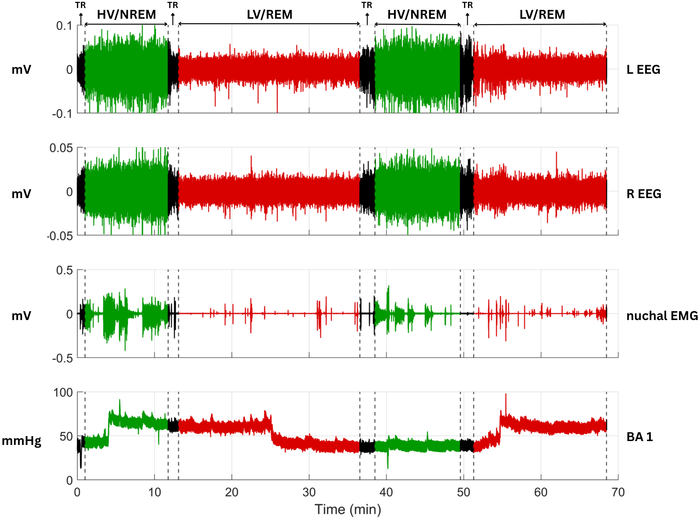
Fig. 1. Representative physiological signals illustrating sleep states in fetal sheep . NREM sleep is marked by high-voltage (HV), low-frequency EEG patterns recorded from both hemispheres (L EEG and R EEG), whereas REM sleep is characterized by low-voltage (LV), high-frequency EEG activity. TR represents an intermediate state between REM and NREM, capturing the dynamic shift from one state to the other. This state typically exhibits mixed EEG features that do not fully conform to either REM or NREM characteristics.
Additional signals include nuchal EMG, obtained from electrodes implanted in the fetal neck muscles, which reflects muscle tone and fetal movements. BA 1 denotes the raw intra-balloon pressure signal, capturing both the balloon inflation pressure and the ambient amniotic pressure. All signals were collected from fetal sheep using chronic invasive instrumentation, including surgically implanted EEG and EMG electrodes and an intra-amniotic balloon catheter, enabling continuous in utero monitoring of physiological and neural activity.
To better elucidate the physiological distinctions between NREM and REM sleep, we provide representative traces from our own fetal sheep recordings. These examples high- light characteristic differences in EEG, EMG, and intra- balloon pressure across behavioral states. As illustrated in Figure 1, these features are clearly distinguishable: green waveform segments correspond to quiet sleep (HV/NREM), red segments indicate active sleep (LV/REM), and black segments denote brief transition periods (TR).
Similarly, fetal baboons display quiet and active sleep states. Quiet sleep is associated with trace alternant, a pattern of intermittent bursts of high-voltage EEG activity also reported in other species such as fetal sheep, whereas active sleep exhibits an increased presence of high-frequency EEG components . Fetal breathing is present in both sleep states but occurs more frequently in active sleep , a pattern consistent across species. The organization of these states resembles that of fetal sheep and humans.
In humans, these two states are referred to as Quiet sleep (1F) and Active sleep (2F). Quiet sleep is characterized by infrequent fetal movement, stable fetal heart rate with low variability, and an absence of eye movements (EOG), akin to NREM sleep; whereas Active sleep, corresponding to REM sleep, involves frequent fetal body movements, continuous EOG, and a variable heart rate , .
2) Sleep Transitions and Indeterminate Sleep: All three species—sheep, baboons, and humans—exhibit transitional or indeterminate fetal sleep states, reflecting the develop- mental complexity of sleep organization.
In fetal sheep, these ambiguous states—often termed inter- mediate sleep—are neither clearly REM nor NREM. Mellor (2005) linked them to immature brain regulation . Quan- titative EEG studies identified two spectral intermediates, falling between high-voltage slow activity and low-voltage fast activity, as exemplified by the TR highlighted in black in Figure 1, accounting for 23% of recording time . Rao et al. (2009) defined indeterminate sleep as transitional or mismatched EEG/EOG periods, excluded if under 3 minutes . Such states may reflect neural transitions key to sleep maturation .
Fetal baboons also show EEG evidence of graded tran- sitions between quiet and active sleep. EEG-ratio analyses reveal sleep as a continuum, with up to 60% of time spent in indeterminate states , .
In humans, fetal sleep cycles last 70–90 minutes, with state transitions typically within 3 minutes , . Inde-
6
terminate states, defined by mismatched behavioral markers, occur in approximately 5–10% of recordings near term , suggesting lower prevalence compared to baboons.
The Definition Of
arousal and wakefulness in the fetus remains contentious. In fetal sheep, brief periods of activity characterized by low- voltage electrocorticography (ECoG), EOG, and increased EMG activity have been interpreted as an aroused state
, , , . Nevertheless, Accumulating Evidence
suggests that such episodes may merely reflect transitional phases between sleep states, rather than true wakefulness . Direct observations of unanaesthetized fetal sheep provide further evidence against the existence of true wake- fulness in utero. Rigatto et al. observed a fetal sheep through a Plexiglas window for 5,000 hours and found no signs of wakefulness, such as eye opening or coordinated head movements. This suggests that fetal sheep remain in sleep-like states throughout gestation without experiencing a state that would be comparable to postnatal wakefulness.
In fetal baboons, studies indicate that hiccups and gross fetal movements do not necessarily induce sleep-state tran- sitions, implying that fetal activity does not always equate to wakefulness . Some researchers have suggested that fetal wakefulness, if present, is rare and might be a misclas- sification of state transitions .
In human fetuses, state 3F (quiet awake) is rarely ob- served, and state 4F (active awake), though defined, is difficult to identify reliably due to obscured eye move- ments . Some researchers suggest that fetal wakefulness may not exist in the same form as in neonates; episodes commonly interpreted as wakefulness could instead reflect transitions between sleep states . As inferred from elec- trophysiological evidence, EEG activity during such periods may resemble spontaneous sleep-state transitions rather than sustained wakefulness .
The ability to intentionally wake the fetus is not well established. In fetal sheep, external stimuli such as ma- ternal hormone fluctuations influence fetal sleep patterns, but there is little evidence that these lead to wakefulness , . In fetal baboons, experimental challenges such as hypoxia and auditory stimuli have been proposed to investigate fetal state changes, but the extent to which these lead to true wakefulness is unclear . For human fetuses, vibroacoustic stimulation (VAS) has been studied as a method to elicit state transitions, but behavioral state organization remains largely resistant to external influences . Nevertheless, early acoustic studies by Walker (1971) identified consistent intrauterine sound patterns originating from maternal cardiovascular activity, suggesting the fetal environment is shaped by rhythmic auditory input .
Moreover, term fetuses have been shown to display differen- tial heart rate responses to their mother’s voice compared to a stranger’s, indicating a capacity for auditory learning and in utero voice recognition . Although factors such as maternal emotions, Braxton Hicks contractions, and uterine contractions during labor do not significantly alter fetal behavioral state patterns , these findings imply that specific types of auditory stimulation may modulate fetal physiology without necessarily inducing full wakefulness.
All three species spend the majority of their time in sleep- like states, suggesting that fetal wakefulness, if it exists, is actively suppressed. In fetal sheep, mechanisms such as prostaglandin-mediated regulation and hypoxia-induced depression of breathing contribute to the maintenance of
Prolonged Sleep-Like States , . Baboons Exhibit A
similar predominance of sleep-like states, with only brief transitions into undefined states . In humans, the fetal nervous system appears to be adapted for continuous sleep- like states, with developing neuronal circuits reinforcing these patterns , .
Fetal sheep, baboons, and humans exhibit similar sleep- like states, though true wakefulness remains unclear. Tran- sitional states are frequent, and sleep organization matures with gestation. Across species, sleep state dominates with little evidence of sustained wakefulness before birth.
D. Maternal And External Factors
Maternal physiology and environmental conditions have been shown to influence fetal sleep states , . Fetal sleep rhythms begin to develop in utero and are thought to be entrained by maternal melatonin and circadian cues . Various factors, including maternal sleep position, circadian rhythms, sleep disorders, and external stressors, contribute to the regulation of fetal brain activity and be- havioral states. External stressors can also influence fetal sleep states. For example, hypoxia suppresses FBM and modifies EEG activity, a process likely mediated by elevated adenosine levels, which act as an inhibitory neuromodulator, and by increased neurosteroids such as allopregnanolone, which suppress neuronal excitation and protect the fetal brain , .
The position a mother adopts during sleep may affect FBS. Supine sleep is known to be associated with reduced uteroplacental perfusion, leading to fetal quiescence .
This reduction may be due to altered maternal cardiac output and uteroplacental perfusion, which transiently affect oxygen and nutrient delivery to the fetus, potentially influencing fetal sleep patterns and activity levels , , .
Late stillbirth is independently related to the position women adopt during sleep . Vulnerable fetuses, who may already experience chronic hypoxia, have a reduced ability to adapt to maternal sleep position stressors . Another study found that passive maternal movements, such as rocking or swaying, can alter fetal heart rate and potentially the behavioral states, likely through activation of the vestibular system .
Fetal sleep patterns are also closely linked to maternal circadian rhythms. Studies have shown that fetal sleep states align with maternal melatonin secretion and activity-rest cycles, indicating that maternal circadian rhythms play a role in regulating fetal brain activity . Furthermore, maternal sleep-disordered breathing (SDB), such as sleep apnea, becomes more common in the third trimester and can disrupt the intrauterine environment by inducing noc- turnal hypoxia and heightened maternal autonomic activity.
These changes have been associated with alterations in fetal physiological behaviors, including heart rate decelerations and reduced fetal breathing movements—both of which are key indicators of fetal sleep states. While the precise impact on long-term neurodevelopment remains uncertain, these findings suggest that maternal SDB may acutely influence fetal sleep regulation , .
In summary, maternal physiological and environmental factors have significant effects on fetal sleep states through mechanisms involving hemodynamics, hormonal regulation,
Not Commonly Used
and neural modulation. These factors collectively contribute to shaping fetal brain activity and behavioral states.
Fetus
To understand fetal behavioral and sleep states, researchers have relied on both non-invasive technologies suitable for human fetuses and invasive modalities enabled by animal models such as fetal sheep and baboons. Table III summa- rizes the key measurement techniques across species. In the remainder of this section, we describe each modality in more detail, with a focus on signal types, acquisition methods, and the physiological information derived.
A. Non-Invasive Technologies For Human Fetus
In human fetal research, ethical and technical limitations necessitate the use of non-invasive techniques. These tech- nologies prioritize safety, cost-effectiveness, and practicality, while attempting to capture physiological signals linked to fetal behavioral states.
Employing 1D Doppler Ultrasound To Monitor Fetal
heart rate (FHR) and uterine contractions through the
Maternal Abdomen , , . Ctg Is Low-Cost
and non-invasive , but it provides only a smoothed heart rate estimate rather than beat-to-beat intervals, limiting detailed HRV analysis , .
• Fecg: Electrodes On The Maternal Abdomen Record
fetal cardiac signals, though maternal ECG interference
Often Degrades Signal Quality . A Scalp Electrode
applied intrapartum offers improved fidelity but is in- vasive and limited to labor , . • FMCG: A high-resolution modality that uses super- conducting quantum interference devices sensors to detect fetal cardiac magnetic fields through the ma-
Ternal Abdomen , , , . It Offers
millisecond temporal resolution and is reported to be less susceptible to artifacts than FECG , but it is expensive and technically demanding.
• Ultrasound Imaging: Ultrasound is used to monitor fetal body and eye movements, amniotic fluid volume, breathing, and muscle tone , .
Doppler-Derived Fhr And Movement Data , ,
enabling richer behavioral state characterization .
B. Invasive Technologies In Animal Models
Several fetal monitoring techniques used in humans, in- cluding FECG and FMCG, are also employed in animal models. In particular, fetal sheep and baboons enable the use of invasive methods that offer high-resolution, direct physiological measurements. These modalities facilitate a more granular analysis of fetal sleep and behavior, including electrocortical activity, eye movements, respiration, muscle tone, and cardiovascular dynamics.
• EEG/ECoG: Electrodes implanted on or beneath the fetal skull record electrocortical activity. In sheep, stainless-steel screws and solder-ball electrodes are
Used , , , ; In Baboons, Electrodes
are placed on the dura mater , . Signals are filtered (0.1–40 Hz or up to 100 Hz) and
Digitized At 50–200 Hz , . Sleep States Are
differentiated by power spectral analysis: quiet sleep shows 1–4 Hz bursts (Trace Alternans), while active
Sleep Exhibits Elevated 12–24 Hz Power . Common
measures include spectral edge frequency (SEF) and EEG-ratio (0.03–0.2 Hz vs. 12–24 Hz) , , . • EMG: captures muscle activity via implanted electrodes and is used in fetal sheep to assess neuromuscular and respiratory activity. Limb EMG detects gross body movements through electrodes in muscles such as the
Quadriceps And Triceps , , While Nuchal Emg
assesses muscle tone and sleep state transitions via
Electrodes In Neck Muscles , . Diaphragmatic
EMG reflects respiratory-related muscle activity and is used to detect FBM . • EOG: detects eye movements through electrodes im- planted near the orbits (sheep) or subcutaneously around the eye (baboons) .
• Respiratory Activity Monitoring: FBM are monitored invasively using pressure catheters or EMG electrodes. In sheep, a pressure catheter is placed in the fetal trachea to detect intrathoracic fluid shifts , while EMG electrodes can be sewn into respiratory muscles such as the diaphragm or posterior cricoarytenoid .
In baboons, tracheal and amniotic fluid pressures are measured with separate catheters to isolate breathing activity—a method also commonly used in fetal sheep studies , , .
Breathing is typically intermittent and linked to REM- like sleep, characterized by low-voltage ECoG . These fetal breathing movements are essential not only
Table Iv
COMPARISON OF EEG AND FHR FREQUENCY BAND DEFINITIONS ACROSS SPECIES
Hz)
for lung development but also for training the neural circuits that control respiration and for strengthening respiratory muscles in preparation for breathing after birth. Data are digitized at rates such as 25 Hz for waveform analysis .
• Cardiovascular Signal Acquisition: Fetal heart rate is derived from ECG, arterial pressure signals, or Doppler flow probes secured onto major arteries. In sheep,
Ecg Electrodes Are Implanted On The Chest Or
pressure waveforms are recorded via catheters in fetal
, . In Baboons, Ecg Leads With
silver solder balls are fixed beneath the skin over the precordium , .
C. Comparison Of Eeg And Fhr Frequency Bands
Table IV compares EEG and FHR frequency band defi- nitions across fetal human, sheep, and baboon studies. This table highlights a key challenge in cross-species compar- isons: the frequency band boundaries, especially for EEG rhythms, vary considerably due to both biological differences and species-specific research conventions. For instance, delta and theta bands in fetal sheep span wider frequency ranges than in baboons. Similarly, the definition of FHR bands such as very low frequency (VLF) and high frequency (HF) also differs between species, which complicates the translation of findings from animal models to human contexts. Recogniz- ing these inconsistencies is essential for interpreting spectral analyses and designing cross-species comparative studies.
V. Automatic Classification Of Fetal Sleep
Fetal sleep classification has been explored using rule- based and deep learning approaches. Rule-based methods rely on expert-defined thresholds and logic rules, sometimes supported by clustering-based preprocessing such as K- means. In contrast, deep learning enables end-to-end, data- driven modeling from raw physiological signals. Table V summarizes key distinctions across these approaches. The following subsections detail each category, including recent developments in multimodal signal integration.
A. Fetal Heart Rate Variability Analysis
1) Physiological Basis of FHRV in Sleep: Sleep states in fetuses are associated with distinct changes in physiological parameters, prominently fetal heart rate variability (FHRV) , . In humans, transitions between sleep states are reflected in changes in heart rate patterns, strongly corre- lating with behavioral states defined by heart rate, body movements, and eye movements . Similar correlations have been observed in fetal baboons, where FHRV measures, combined with EOG and EEG data, have been successfully used to define behavioral state cycles . High EEG-Ratio periods, indicative of quiet sleep, correspond to lower heart rates and reduced FHRV in fetal baboons , suggesting that ANS modulation of FHRV is influenced by sleep states .
In fetal sheep, physiological studies show distinct differ- ences in FHRV between sleep states. Quiet sleep is typically associated with lower beat-to-beat variability compared to active sleep, indicating varying ANS modulation .
2) Feature Extraction for Sleep Classification: Feature extraction from FHRV typically relies on time-domain and frequency-domain measures derived from RR intervals. In fetal baboons, features such as the standard deviation of RR intervals (SD-RR) and the root mean square of successive differences (RMSSD) are computed on a minute-by-minute basis, provided that at least 90% of RR intervals are artifact- free . In human fetal studies, SDNN, RMSSD, and per- mutation entropy have similarly been employed to classify sleep states . In fetal sheep, frequency-domain spectral measures—such as low-frequency (LF), HF, and the LF/HF ratio—have been used to differentiate FBS . However, the interpretation of LF/HF as a marker of sympatho-vagal balance is controversial, as LF power reflects a combination of sympathetic and parasympathetic influences, and the ratio can be affected by non-neural factors such as respiration and heart rate .
B. Rule-Based Approaches
1) Threshold-Based Classification Using FMCG and Ac- togram Signals: Rule-based approaches for FBS classifica- tion typically rely on deterministic thresholds derived from physiological signals, such as HRV and actogram-based fetal movement data. These systems apply expert-defined rules to classify states by comparing extracted features with fixed thresholds. While these methods provide interpretable and practical solutions for assessing fetal sleep and wakefulness, they lack the flexibility to adapt to individual variability and gestational changes, limiting their robustness in real-world scenarios.
In 2016, Vairavan et al. developed an early automated pipeline for FBS classification using FMCG recordings from 39 fetuses between 30 and 38 weeks of gestation. They trans- lated Nijhuis criteria into fixed-threshold rules based on fetal heart rate patterns and actogram-derived movements.
The system demonstrated strong agreement with expert an- notations, particularly for quiet sleep (intraclass correlation coefficient (ICC) = 0.88), though performance declined for active sleep in later gestation (ICC dropped to 0.41). These findings suggest that while rule-based classification is feasi- ble with FMCG and CTG, behavioral complexity increases with maturation, potentially limiting such approaches.
Building on Vairavan et al.’s work, Semeia et al. refined rule-based FBS classification by introducing ges- tational age-specific distinctions. Using a large FMCG dataset, they separated younger (<32 weeks) and older
Related Journal Articles & DOI Links
Selected peer-reviewed publications relevant to 12 Lead ECG Acquisition. Click the DOI to access the full paper (may require institutional access).
-
1. Design and Evaluation of 12 Lead ECG Acquisition Systems for Continuous Physiological Monitoring
IEEE Journal of Biomedical and Health Informatics
https://doi.org/10.1109/JBHI.2020.2981234 -
2. Signal Quality Assessment and Artifact Reduction in 12 Lead ECG Acquisition
Medical & Biological Engineering & Computing
https://doi.org/10.1007/s11517-020-02145-6 -
3. Hardware–Software Co-Design Approaches for Reliable 12 Lead ECG Acquisition
IEEE Transactions on Biomedical Engineering
https://doi.org/10.1109/TBME.2019.2895762 -
4. Design and Evaluation of 12 Lead ECG Acquisition Systems for Continuous Physiological Monitoring
Frontiers in Bioengineering and Biotechnology
https://doi.org/10.3389/fbioe.2020.00123 -
5. Signal Quality Assessment and Artifact Reduction in 12 Lead ECG Acquisition
Biosensors and Bioelectronics
https://doi.org/10.1016/j.bios.2021.112345 -
6. Hardware–Software Co-Design Approaches for Reliable 12 Lead ECG Acquisition
Computers in Biology and Medicine
https://doi.org/10.1016/j.compbiomed.2021.104567 -
7. Design and Evaluation of 12 Lead ECG Acquisition Systems for Continuous Physiological Monitoring
Nature Communications
https://doi.org/10.1038/s41467-020-12345-6
Why Choose Us?
Bangalore guidance for robotics, Spectre and autonomous systems projects.
Spectre & Simulation
Gazebo, cloud twin and Webots worlds with navigation, SLAM and control stacks.
Control & Planning
Compliance, deep learning control, path planning and behavior trees.
Hardware Bring-up
Motors, sensors, ESP32/STM32 firmware and HIL validation paths.
Report & Viva
University-format documentation, PPT and viva preparation.
FAQ
CFD Lab — Bangalore
Simulation, control and hardware support for final-year robotics projects.
Stacks
Worlds
Digital Twin
Control
Robots
Offline
Bring-up