Towards Electrophysiological and Histological Mapping of Upper
Limb Nerves In Pigs Using Epineural Stimulation
Baum J1, Chamot-Nonin M2, Oppelt V3, Guiraud D2, Azevedo Coste C1, Guiho T1
3Cortec Company, Freiburg, Germany
Abstract: Understanding the relationship between nerve anatomy and the functional outcomes of electrical stimulation is critical for optimizing neural interface design. In this study, we conducted acute experiments on four pigs in which epineural cuff electrodes with multiple contacts were placed around upper limb nerves. A subset of electrical stimulation configurations - previously identified via computational study - was applied, and the resulting evoked electromyographic (EMG) responses were recorded from target muscles. Muscle recruitment curves were extracted and analysed offline to quantify activation patterns. Following the electrophysiological experiments, the stimulated nerves were harvested and processed for histological analysis to visualize fascicular organization and distribution. This work presents preliminary results from the combined analysis of muscle activation profiles and fascicle anatomy in one animal. Our findings aim to inform the design of stimulation strategies by linking electrode configuration to selective muscle recruitment, ultimately contributing to more effective neuromodulation and neuroprosthetic applications.
Keywords: epineural stimulation, swine, needle EMG, histology, selectivity
Introduction
Implanted peripheral nerve stimulation has shown promise in restoring impaired motor functions of the upper limb— particularly hand and wrist movements—in individuals
With Complete Spinal Cord Injury . Compared To
epimysial or intraneural electrodes, the use of multicontact epineural electrodes offers a less invasive alternative while maintaining access to multiple motor pathways .
However, achieving selective activation of specific muscle subgroups to produce functional movements remains a significant challenge. Currently, stimulation parameters and electrode configuration are typically adjusted empirically, based on observed motor responses, which limits reproducibility and precision. This approach is further complicated by inter- and intraindividual variability in nerve morphology, particularly regarding the complex fascicular organization and plexiform architecture of the
Human Brachial Plexus . Understanding How These
anatomical factors influence stimulation outcomes is essential for optimizing electrode design and stimulation strategies.
To partially address this scientific challenge, equivalent electrical models of axons were developed to enable the in- silico identification of the most selective and efficient current configurations. Cuff epineural electrodes were considered in the model due to their moderately invasive characteristics and widespread commercial availability - in contrast to other architectures such as TIME intraneural electrodes which might be less suitable for clinical trials due to mechanical constraints associated with their transversal positioning through the nerves. Previously, subset of stimulation configurations identified as selective by the model was evaluated in 28-days clinical trials investigating peripheral nerve stimulation for hand and wrist rehabilitation . These configurations were further
Refined Empirically Using Evoked Electromyography
(eEMG) and observation of evoked movements to elicit specific functional gestures. However, this empirical approach is time-consuming, and only a limited portion of the extensive parameter space can be explored clinically for the refinement of epineural functional electrical stimulation (FES). Optimization of stimulation parameters
More
comprehensive and objective, while remaining time- realistic. The development of automated tools capable of correlating epineural current patterns with evoked electromyographic (eEMG) responses is essential to systematically explore the extensive parameter space and enhance individualized epineural stimulation. However,
Existing Models Are Either Based On Stereotypical
assumptions of nerve structure , or on histological observations that lack electrophysiological validation . A recent study attempted to bridge this gap by combining anatomical and electrophysiological data using a specific epineural electrode , but the stimulation paradigm was limited to bipolar configurations. While this work highlights the value of building subject-specific models, it remains constrained in selectivity. This challenge is then compounded by inter-individual variability, which further complicates the development of accurate and generalizable models. With these considerations in mind, we designed a preclinical study that aimed at elucidating the relationship
And
electrophysiologically evoked responses. To enhance the translational relevance of the findings, priority was given to a large animal species. In particular, the selected species – the domestic pig (sus scrofa domesticus) - exhibits
Anatomical Similarities To Humans, Notably In The
distribution of the brachial plexus, including the median, radial, and ulnar nerves, which are functionally conserved in controlling wrist and finger flexion and extension. .
In this article, on top of confirming the relevance of swine for electrophysiological studies of upper limbs functions, we exploited eEMGs to draw a parallel between evoked muscular activities elicited by a subset of presumably selective multipolar stimulation configuration/parameters and actual nerve anatomy - obtained via histological analyses of stimulated nerves.
Number
#47593-2024021614231926v2), acute pre-clinical trials were conducted on four neurotypical juvenal swine (2-3 months age, 30-40 kg).
After Premedication, Anaesthesia Was Induced With
propofol and maintained via intravenous infusion of propofol mixed with sufentanil for analgesia. Swine condition was then monitored throughout the procedure (body temperature; heart rate; blood saturation; breathing rate with air and oxygen mechanically assisted ventilation).
After ensuring proper anaesthesia, swine axillary fossa was incised to expose the brachial plexus. After anatomical prospection, the median nerve was further identified with a bipolar stimulation stick inducing contraction of
Predefined Forelimb Muscles, Namely “Flexor Carpi
radialis” (FCR), “Flexor digitorum superficialis” (FDS), "Pronator teres" (PT). “Extensor carpi radialis” (ECR) muscle was also identified to enable monitoring of antagonist contractions. A multichannel cuff electrode (CorTec™, Germany) with two plain contacts on distal and
Contacts
distributed along the central circumference (Fig. 1, see for further details) was then wrapped around the nerve before connection to a 16 channels bench-top neural
For
electrophysiological measurements. Armpit incision was then filled with saline to enable moisturization of the nerve and electrical continuity between the electrode contacts and the nerve.
Protocol
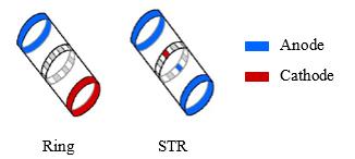
Epineural stimulation was delivered using asymmetric biphasic pulses - 150 µs phase width - at a frequency of 35 Hz. Model-based preidentified stimulation configurations were investigated, ie. rings and “steering current” (STR).
The STR configuration is characterized by the distribution of current between one cathode and three anodes leading to anodic current repartition of 1/3 on each ring and on the inner contact opposite to the cathodic one. STR are numbered from 1 to 6 according to cathodic central contact position around nerve circumference (Fig. 1, ).
For each configuration (7 in total), stimulation intensity was initially set at a low value (few tens of µV) before gradually increasing - step of 9 µA - until estimation of saturation of the muscle recruitment. Each step duration was 4.5 s long leading to trains of 19 stimulation pulses and 19 M-waves per intensity.
Figure 1: Schematic representation of ring and STR
Configurations. Adapted From
Targeted muscles were localized via skin palpation further confirmed through stimulation-evoked contractions and movements. Pairs of Teflon-insulated stainless-steel wires were inserted percutaneously to record evoked EMGs (eEMGs). The signals were then amplified with a gain of
5000, Band Pass Filtered Between 1 Hz And 5 Khz
(Butterworth filter of 4th order) et notch filtered at 50 Hz
(Powerlab 16/35, Ad Instrument, New Zealand). The
associated LabChart software (LabChart 7, AD Instrument, New Zealand) was used throughout the experiments to
Histological Protocol
Following euthanasia of the swines, both the stimulated
And Control Nerves Were Harvested And Immediately
immersed in saline solution. The positions of the electrode contacts were marked on the epineurium using tattoo ink prior to nerve sectioning. Nerve fragments were fixed in 10% paraformaldehyde (PFA) for 48 hours, followed by dehydration through a graded ethanol series prior to
Paraffin Embedding. The Paraffin-Embedded Nerve
fragments were sectioned at a thickness of 3 µm and subsequently stained with hematoxylin-eosin-saffron (HES) to visualize intraneural structures, or processed for choline acetyltransferase (ChAT) immunohistochemistry to specifically evaluate the distribution of motor fibers within the fascicles.
Eemgs Processing
Raw recorded eEMGs were further band pass filtered between 10 and 500 Hz (Butterworth filter of 4th order). For each muscle and each stimulation intensity, repeated M-waves were averaged before peak-to-peak calculation.
The obtained data were then normalized using maximum eEMG peak-to-peak values (obtained over all recordings) before plotting recruitment curves.
Results
In this section only preliminary results from pig #2 (P#2) are presented.
An
electrophysiological study of upper limbs innervation.
Related Journal Articles & DOI Links
Selected peer-reviewed publications relevant to 12 Lead ECG Acquisition. Click the DOI to access the full paper (may require institutional access).
-
1. Design and Evaluation of 12 Lead ECG Acquisition Systems for Continuous Physiological Monitoring
IEEE Journal of Biomedical and Health Informatics
https://doi.org/10.1109/JBHI.2020.2981234 -
2. Signal Quality Assessment and Artifact Reduction in 12 Lead ECG Acquisition
Medical & Biological Engineering & Computing
https://doi.org/10.1007/s11517-020-02145-6 -
3. Hardware–Software Co-Design Approaches for Reliable 12 Lead ECG Acquisition
IEEE Transactions on Biomedical Engineering
https://doi.org/10.1109/TBME.2019.2895762 -
4. Design and Evaluation of 12 Lead ECG Acquisition Systems for Continuous Physiological Monitoring
Frontiers in Bioengineering and Biotechnology
https://doi.org/10.3389/fbioe.2020.00123 -
5. Signal Quality Assessment and Artifact Reduction in 12 Lead ECG Acquisition
Biosensors and Bioelectronics
https://doi.org/10.1016/j.bios.2021.112345 -
6. Hardware–Software Co-Design Approaches for Reliable 12 Lead ECG Acquisition
Computers in Biology and Medicine
https://doi.org/10.1016/j.compbiomed.2021.104567 -
7. Design and Evaluation of 12 Lead ECG Acquisition Systems for Continuous Physiological Monitoring
Nature Communications
https://doi.org/10.1038/s41467-020-12345-6
Why Choose Us?
Bangalore guidance for robotics, Spectre and autonomous systems projects.
Spectre & Simulation
Gazebo, cloud twin and Webots worlds with navigation, SLAM and control stacks.
Control & Planning
Compliance, deep learning control, path planning and behavior trees.
Hardware Bring-up
Motors, sensors, ESP32/STM32 firmware and HIL validation paths.
Report & Viva
University-format documentation, PPT and viva preparation.
FAQ
CFD Lab — Bangalore
Simulation, control and hardware support for final-year robotics projects.
Stacks
Worlds
Digital Twin
Control
Robots
Offline
Bring-up